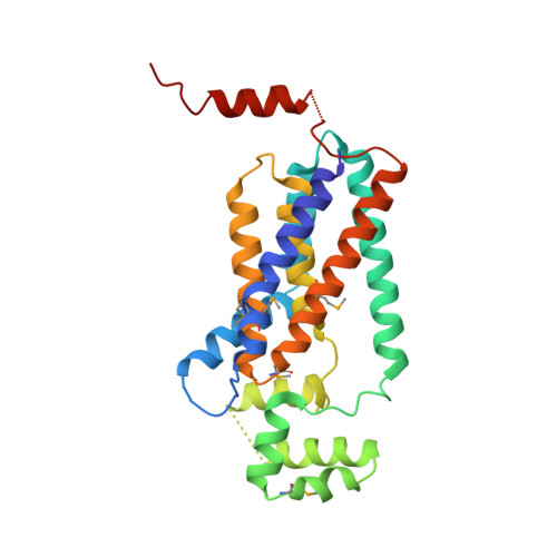
4PX7

Crystal structure of lipid phosphatase E. coli PgpB


Experimental Data Snapshot

  • Method: X-RAY DIFFRACTION
  • Resolution: 3.20 Å
  • R-Value Free: 0.302 
  • R-Value Work: 0.270 
  • R-Value Observed: 0.272 

wwPDB Validation   3D Report Full Report


Ligand Structure Quality Assessment 


This is version 1.1 of the entry. See complete history


Literature

Crystal structure of lipid phosphatase Escherichia coli phosphatidylglycerophosphate phosphatase B.

Fan, J.Jiang, D.Zhao, Y.Liu, J.Zhang, X.C.

(2014) Proc Natl Acad Sci U S A 111: 7636-7640

  • DOI: https://doi.org/10.1073/pnas.1403097111
  • Primary Citation of Related Structures:  
    4PX7

  • PubMed Abstract: 

    Membrane-integrated type II phosphatidic acid phosphatases (PAP2s) are important for numerous bacterial to human biological processes, including glucose transport, lipid metabolism, and signaling. Escherichia coli phosphatidylglycerol-phosphate phosphatase B (ecPgpB) catalyzes removing the terminal phosphate group from a lipid carrier, undecaprenyl pyrophosphate, and is essential for transport of many hydrophilic small molecules across the membrane. We determined the crystal structure of ecPgpB at a resolution of 3.2 Å. This structure shares a similar folding topology and a nearly identical active site with soluble PAP2 enzymes. However, the substrate binding mechanism appears to be fundamentally different from that in soluble PAP2 enzymes. In ecPgpB, the potential substrate entrance to the active site is located in a cleft formed by a V-shaped transmembrane helix pair, allowing lateral movement of the lipid substrate entering the active site from the membrane lipid bilayer. Activity assays of point mutations confirmed the importance of the catalytic residues and potential residues involved in phosphate binding. The structure also suggests an induced-fit mechanism for the substrate binding. The 3D structure of ecPgpB serves as a prototype to study eukaryotic PAP2 enzymes, including human glucose-6-phosphatase, a key enzyme in the homeostatic regulation of blood glucose concentrations.


  • Organizational Affiliation

    National Laboratory of Macromolecules, National Center of Protein Science-Beijing, Institute of Biophysics, Chinese Academy of Sciences, Beijing 100101, China;Graduate School of the University of Chinese Academy of Sciences, Beijing 100049, China;


Macromolecules
Find similar proteins by:  (by identity cutoff)  |  3D Structure
Entity ID: 1
MoleculeChains Sequence LengthOrganismDetailsImage
Phosphatidylglycerophosphatase262Escherichia coli BL21(DE3)Mutation(s): 2 
Gene Names: pgpBB21_01266ECBD_2341ECD_01255
EC: 3.1.3.27 (PDB Primary Data), 3.6.1.27 (PDB Primary Data)
Entity Groups  
Sequence Clusters30% Identity50% Identity70% Identity90% Identity95% Identity100% Identity
Sequence Annotations
Expand
  • Reference Sequence
Small Molecules
Ligands 2 Unique
IDChains Name / Formula / InChI Key2D Diagram3D Interactions
LDA
Query on LDA

Download Ideal Coordinates CCD File 
B [auth A]LAURYL DIMETHYLAMINE-N-OXIDE
C14 H31 N O
SYELZBGXAIXKHU-UHFFFAOYSA-N
GOL
Query on GOL

Download Ideal Coordinates CCD File 
C [auth A]GLYCEROL
C3 H8 O3
PEDCQBHIVMGVHV-UHFFFAOYSA-N
Modified Residues  1 Unique
IDChains TypeFormula2D DiagramParent
MSE
Query on MSE
A
L-PEPTIDE LINKINGC5 H11 N O2 SeMET
Experimental Data & Validation

Experimental Data

  • Method: X-RAY DIFFRACTION
  • Resolution: 3.20 Å
  • R-Value Free: 0.302 
  • R-Value Work: 0.270 
  • R-Value Observed: 0.272 
  • Space Group: P 21 21 21
Unit Cell:
Length ( Å )Angle ( ˚ )
a = 36.715α = 90
b = 73.667β = 90
c = 131.2γ = 90
Software Package:
Software NamePurpose
HKL-2000data collection
SOLVEphasing
PHENIXrefinement
DENZOdata reduction
SCALAdata scaling

Structure Validation

View Full Validation Report


Ligand Structure Quality Assessment 


Entry History 

Deposition Data

Revision History  (Full details and data files)

  • Version 1.0: 2014-05-28
    Type: Initial release
  • Version 1.1: 2022-08-24
    Changes: Database references, Derived calculations