Structural basis for 18-beta-glycyrrhetinic acid as a novel non-GSH analog glyoxalase I inhibitor
Zhang, H., Huang, Q., Zhai, J., Zhao, Y.N., Zhang, L.P., Chen, Y.Y., Zhang, R.W., Li, Q., Hu, X.P.(2015) Acta Pharmacol Sin 36: 1145-1150
- PubMed: 26279158 Search on PubMedSearch on PubMed Central
- DOI: https://doi.org/10.1038/aps.2015.59
- Primary Citation Related Structures:
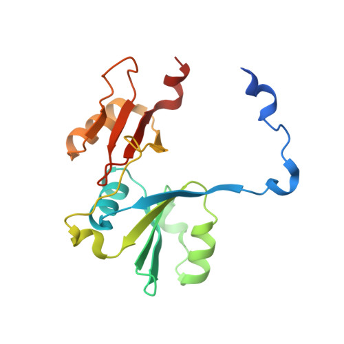
4PV5 - PubMed Abstract:
Glyoxalase I (GLOI), a glutathione (GSH)-dependent enzyme, is overexpressed in tumor cells and related to multi-drug resistance in chemotherapy, making GLOI inhibitors as potential anti-tumor agents. But the most studied GSH analogs exhibit poor pharmacokinetic properties. The aim of this study was to discover novel non-GSH analog GLOI inhibitors and analyze their binding mechanisms. Mouse GLOI (mGLOI) was expressed in BL21 (DE3) pLysS after induction with isopropyl-β-D-1-thiogalactopyranoside and purified using AKTA FPLC system. An in vitro mGLOI enzyme assay was used to screen a small pool of compounds containing carboxyl groups. Crystal structure of the mGLOI-inhibitor complex was determined at 2.3 Å resolution. Molecular docking study was performed using Discovery Studio 2.5 software package. A natural compound 18-β-glycyrrhetinic acid (GA) and its derivative carbenoxolone were identified as potent competitive non-GSH analog mGLOI inhibitors with Ki values of 0.29 μmol/L and 0.93 μmol/L, respectively. Four pentacyclic triterpenes (ursolic acid, oleanolic acid, betulic acid and tripterine) showed weak activities (mGLOI inhibition ratio <25% at 10 μmol/L) and other three (maslinic acid, corosolic acid and madecassic acid) were inactive. The crystal structure of the mGLOI-GA complex showed that the carboxyl group of GA mimicked the γ-glutamyl residue of GSH by hydrogen bonding to the glutamyl sites (residues Arg38B, Asn104B and Arg123A) in the GSH binding site of mGLOI. The extensive van der Waals interactions between GA and the surrounding residues also contributed greatly to the binding of GA and mGLOI. This work demonstrates a carboxyl group to be an important functional feature of non-GSH analog GLOI inhibitors.
- School of Pharmaceutical Sciences & Centre for Cellular and Structural Biology of Sun Yat-sen University, Guangzhou 510006, China.
Organizational Affiliation: