4PPO | pdb_00004ppo

First Crystal Structure for an Oxaliplatin-Protein Complex


Experimental Data Snapshot

  • Method: X-RAY DIFFRACTION
  • Resolution: 1.73 Å
  • R-Value Free: 
    0.235 (Depositor), 0.233 (DCC) 
  • R-Value Work: 
    0.180 (Depositor), 0.191 (DCC) 
  • R-Value Observed: 
    0.181 (Depositor) 

Starting Model: experimental
View more details

wwPDB Validation 3D Report Full Report

Validation slider image for 4PPO

Ligand Structure Quality Assessment 


This is version 1.3 of the entry. See complete history

Literature

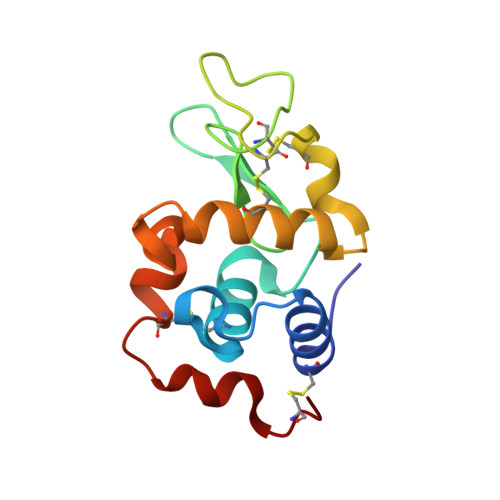
The X-ray structure of the complex formed in the reaction between oxaliplatin and lysozyme.

Messori, L.Marzo, T.Merlino, A.

(2014) Chem Commun (Camb) 50: 8360-8362

  • DOI: https://doi.org/10.1039/c4cc02254h
  • Primary Citation Related Structures: 
    4PPO

  • PubMed Abstract: 

    The X-ray structure of the adduct formed between oxaliplatin and the model protein hen egg white lysozyme is reported here. The structure is compared with those of cisplatin and carboplatin derivatives, previously solved. Relevant changes are highlighted among these crystal structures that are suggestive of significant differences in the reactivity of platinum drugs with this protein; possible biological implications are discussed.


  • Organizational Affiliation
    • Department of Chemistry, University of Florence, Via della Lastruccia 3, 50019 Sesto Fiorentino (FI), Italy.

Macromolecule Content 

  • Total Structure Weight: 15.2 kDa 
  • Atom Count: 1,163 
  • Modeled Residue Count: 129 
  • Deposited Residue Count: 129 
  • Unique protein chains: 1

Macromolecules

Find similar proteins by:|  3D Structure
Entity ID: 1
MoleculeChains  Sequence LengthOrganismDetailsImage
Lysozyme C129Gallus gallusMutation(s): 0 
Gene Names: LYZ
EC: 3.2.1.17
UniProt
Find proteins for P00698 (Gallus gallus)
Explore P00698 
Go to UniProtKB:  P00698
Entity Groups
Sequence Clusters30% Identity50% Identity70% Identity90% Identity95% Identity100% Identity
UniProt GroupP00698
Sequence Annotations
Expand
Reference Sequence

Small Molecules

Ligands 3 Unique
IDChains Name / Formula / InChI Key2D Diagram3D Interactions
1PT

Query on 1PT


Download:Ideal Coordinates CCD File
K [auth A]CYCLOHEXANE-1(R),2(R)-DIAMINE-PLATINUM(II)
C6 H14 N2 Pt
SGLJYTWMWIAGEU-KGZKBUQUSA-N
EDO

Query on EDO


Download:Ideal Coordinates CCD File
B [auth A],
C [auth A]
1,2-ETHANEDIOL
C2 H6 O2
LYCAIKOWRPUZTN-UHFFFAOYSA-N
NO3

Query on NO3


Download:Ideal Coordinates CCD File
D [auth A]
E [auth A]
F [auth A]
G [auth A]
H [auth A]
D [auth A],
E [auth A],
F [auth A],
G [auth A],
H [auth A],
I [auth A],
J [auth A]
NITRATE ION
N O3
NHNBFGGVMKEFGY-UHFFFAOYSA-N

Experimental Data & Validation

Experimental Data

  • Method: X-RAY DIFFRACTION
  • Resolution: 1.73 Å
  • R-Value Free:  0.235 (Depositor), 0.233 (DCC) 
  • R-Value Work:  0.180 (Depositor), 0.191 (DCC) 
  • R-Value Observed: 0.181 (Depositor) 
Space Group: P 43 21 2
Unit Cell:
Length ( Å )Angle ( ˚ )
a = 77.049α = 90
b = 77.049β = 90
c = 37.287γ = 90
Software Package:
Software NamePurpose
REFMACrefinement
CNSrefinement
CrystalCleardata collection
HKL-2000data reduction
HKL-2000data scaling
CNSphasing

Structure Validation

View Full Validation Report


Ligand Structure Quality Assessment 


Entry History 

Deposition Data

Revision History  (Full details and data files)

  • Version 1.0: 2015-01-14
    Type: Initial release
  • Version 1.1: 2019-07-17
    Changes: Data collection, Refinement description
  • Version 1.2: 2023-09-20
    Changes: Data collection, Database references, Derived calculations, Refinement description
  • Version 1.3: 2024-10-30
    Changes: Structure summary