4PG9

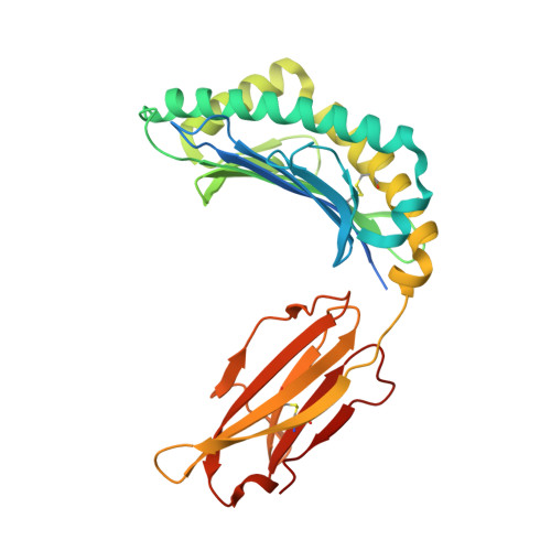
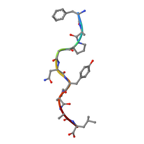
MHC Class I in complex with Sendai virus nucleoprotein peptide FAPGNYPAL


Experimental Data Snapshot

  • Method: X-RAY DIFFRACTION
  • Resolution: 2.40 Å
  • R-Value Free: 0.250 
  • R-Value Work: 0.210 
  • R-Value Observed: 0.212 

Starting Model: experimental
View more details

wwPDB Validation   3D Report Full Report


This is version 1.7 of the entry. See complete history


Literature

The first step of peptide selection in antigen presentation by MHC class I molecules.

Garstka, M.A.Fish, A.Celie, P.H.Joosten, R.P.Janssen, G.M.Berlin, I.Hoppes, R.Stadnik, M.Janssen, L.Ovaa, H.van Veelen, P.A.Perrakis, A.Neefjes, J.

(2015) Proc Natl Acad Sci U S A 112: 1505-1510

  • DOI: https://doi.org/10.1073/pnas.1416543112
  • Primary Citation of Related Structures:  
    4PG9, 4PGB, 4PGC, 4PGD, 4PGE

  • PubMed Abstract: 

    MHC class I molecules present a variable but limited repertoire of antigenic peptides for T-cell recognition. Understanding how peptide selection is achieved requires mechanistic insights into the interactions between the MHC I and candidate peptides. We find that, at first encounter, MHC I H-2K(b) considers a wide range of peptides, including those with expanded N termini and unfitting anchor residues. Discrimination occurs in the second step, when noncanonical peptides dissociate with faster exchange rates. This second step exhibits remarkable temperature sensitivity, as illustrated by numerous noncanonical peptides presented by H-2K(b) in cells cultured at 26 °C relative to 37 °C. Crystallographic analyses of H-2K(b)-peptide complexes suggest that a conformational adaptation of H-2K(b) drives the decisive step in peptide selection. We propose that MHC class I molecules consider initially a large peptide pool, subsequently refined by a temperature-sensitive induced-fit mechanism to retain the canonical peptide repertoire.


  • Organizational Affiliation

    Departments of Cell Biology and.


Macromolecules
Find similar proteins by:  (by identity cutoff)  |  3D Structure
Entity ID: 1
MoleculeChains Sequence LengthOrganismDetailsImage
H-2 class I histocompatibility antigen, K-B alpha chain304Mus musculusMutation(s): 0 
Gene Names: H2-K1H2-K
UniProt
Find proteins for P01901 (Mus musculus)
Explore P01901 
Go to UniProtKB:  P01901
Entity Groups  
Sequence Clusters30% Identity50% Identity70% Identity90% Identity95% Identity100% Identity
UniProt GroupP01901
Sequence Annotations
Expand
  • Reference Sequence
Find similar proteins by:  (by identity cutoff)  |  3D Structure
Entity ID: 2
MoleculeChains Sequence LengthOrganismDetailsImage
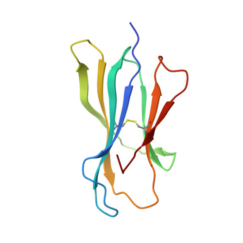
Beta-2-microglobulin100Mus musculusMutation(s): 1 
Gene Names: B2m
UniProt & NIH Common Fund Data Resources
Find proteins for P01887 (Mus musculus)
Explore P01887 
Go to UniProtKB:  P01887
IMPC:  MGI:88127
Entity Groups  
Sequence Clusters30% Identity50% Identity70% Identity90% Identity95% Identity100% Identity
UniProt GroupP01887
Sequence Annotations
Expand
  • Reference Sequence

Find similar proteins by:  Sequence   |   3D Structure  

Entity ID: 3
MoleculeChains Sequence LengthOrganismDetailsImage
Sendai virus nucleoprotein9Respirovirus murisMutation(s): 0 
UniProt
Find proteins for O57286 (Sendai virus (strain Ohita))
Explore O57286 
Go to UniProtKB:  O57286
Entity Groups  
UniProt GroupO57286
Sequence Annotations
Expand
  • Reference Sequence
Small Molecules
Ligands 1 Unique
IDChains Name / Formula / InChI Key2D Diagram3D Interactions
GOL
Query on GOL

Download Ideal Coordinates CCD File 
D [auth A]
E [auth A]
F [auth A]
G [auth A]
H [auth A]
D [auth A],
E [auth A],
F [auth A],
G [auth A],
H [auth A],
I [auth B],
J [auth B],
K [auth B],
L [auth B],
M [auth C]
GLYCEROL
C3 H8 O3
PEDCQBHIVMGVHV-UHFFFAOYSA-N
Experimental Data & Validation

Experimental Data

  • Method: X-RAY DIFFRACTION
  • Resolution: 2.40 Å
  • R-Value Free: 0.250 
  • R-Value Work: 0.210 
  • R-Value Observed: 0.212 
  • Space Group: P 21 21 2
Unit Cell:
Length ( Å )Angle ( ˚ )
a = 87.721α = 90
b = 136.533β = 90
c = 45.203γ = 90
Software Package:
Software NamePurpose
SCALAdata scaling
XDSdata scaling
PDB_EXTRACTdata extraction
PHASERphasing
REFMACrefinement

Structure Validation

View Full Validation Report


Entry History & Funding Information

Deposition Data


Funding OrganizationLocationGrant Number
Netherlands Organisation for Scientific Research (NWO)Netherlands--

Revision History  (Full details and data files)

  • Version 1.0: 2015-01-07
    Type: Initial release
  • Version 1.1: 2015-01-21
    Changes: Database references
  • Version 1.2: 2015-02-04
    Changes: Database references
  • Version 1.3: 2015-02-11
    Changes: Database references
  • Version 1.4: 2017-11-22
    Changes: Advisory, Database references, Derived calculations, Other, Refinement description, Source and taxonomy
  • Version 1.5: 2020-01-08
    Changes: Author supporting evidence
  • Version 1.6: 2023-09-27
    Changes: Data collection, Database references, Refinement description
  • Version 1.7: 2024-10-23
    Changes: Structure summary