


Crystal structure of M. tuberculosis in complex with a cBT
Neres, J., Cole, S.To be published.
Experimental Data Snapshot
Entity ID: 1 | |||||
|---|---|---|---|---|---|
| Molecule | Chains | Sequence Length | Organism | Details | Image |
| Probable decaprenylphosphoryl-beta-D-ribose oxidase | 480 | Mycobacterium tuberculosis H37Rv | Mutation(s): 0 Gene Names: dprE1, Rv3790 EC: 1 (PDB Primary Data), 1.1.98.3 (UniProt) | 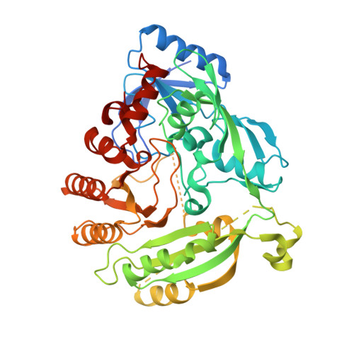 | |
UniProt | |||||
Entity Groups | |||||
| Sequence Clusters | 30% Identity50% Identity70% Identity90% Identity95% Identity100% Identity | ||||
| UniProt Group | P9WJF1 | ||||
Sequence AnnotationsExpand | |||||
Reference Sequence | |||||
| Ligands 3 Unique | |||||
|---|---|---|---|---|---|
| ID | Chains | Name / Formula / InChI Key | 2D Diagram | 3D Interactions | |
| FAD Download:Ideal Coordinates CCD File | C [auth A], F [auth B] | FLAVIN-ADENINE DINUCLEOTIDE C27 H33 N9 O15 P2 VWWQXMAJTJZDQX-UYBVJOGSSA-N |  | ||
| 2R2 Download:Ideal Coordinates CCD File | E [auth A], G [auth B] | [4-(2-methoxyethyl)piperazin-1-yl][6-methyl-7-nitro-5-(trifluoromethyl)-1,3-benzothiazol-2-yl]methanone C17 H19 F3 N4 O4 S AOMUWGOWJLARPE-UHFFFAOYSA-N |  | ||
| IMD Download:Ideal Coordinates CCD File | D [auth A], H [auth B] | IMIDAZOLE C3 H5 N2 RAXXELZNTBOGNW-UHFFFAOYSA-O |  | ||
| Length ( Å ) | Angle ( ˚ ) |
|---|---|
| a = 59.521 | α = 90 |
| b = 84.129 | β = 100.73 |
| c = 90.149 | γ = 90 |
| Software Name | Purpose |
|---|---|
| XDS | data scaling |
| PHASER | phasing |
| REFMAC | refinement |
| Funding Organization | Location | Grant Number |
|---|---|---|
| European Commission | MM4TB - 260872 |


