


High Resolution Structural Views of Rubidium, Cesium and Barium Binding within a Potassium Selective Channel Filter
Lam, Y., Zeng, W., Sauer, D.B., Jiang, Y.To be published.
Experimental Data Snapshot
wwPDB Validation 3D Report Full Report

Entity ID: 1 | |||||
|---|---|---|---|---|---|
| Molecule | Chains | Sequence Length | Organism | Details | Image |
| Potassium channel protein | 97 | Bacillus cereus ATCC 14579 | Mutation(s): 2 Gene Names: BC_0669 Membrane Entity: Yes | 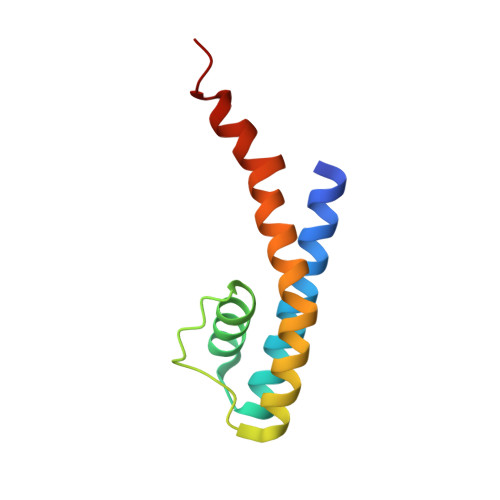 | |
UniProt | |||||
Entity Groups | |||||
| Sequence Clusters | 30% Identity50% Identity70% Identity90% Identity95% Identity100% Identity | ||||
| UniProt Group | Q81HW2 | ||||
Sequence AnnotationsExpand | |||||
Reference Sequence | |||||
| Ligands 4 Unique | |||||
|---|---|---|---|---|---|
| ID | Chains | Name / Formula / InChI Key | 2D Diagram | 3D Interactions | |
| CS Download:Ideal Coordinates CCD File | C [auth A], D [auth A], E [auth A], J [auth B], K [auth B] | CESIUM ION Cs NCMHKCKGHRPLCM-UHFFFAOYSA-N |  | ||
| MPD Download:Ideal Coordinates CCD File | G [auth A], H [auth A], L [auth B] | (4S)-2-METHYL-2,4-PENTANEDIOL C6 H14 O2 SVTBMSDMJJWYQN-YFKPBYRVSA-N |  | ||
| HEX Download:Ideal Coordinates CCD File | I [auth A], M [auth B] | HEXANE C6 H14 VLKZOEOYAKHREP-UHFFFAOYSA-N |  | ||
| NA Download:Ideal Coordinates CCD File | F [auth A] | SODIUM ION Na FKNQFGJONOIPTF-UHFFFAOYSA-N |  | ||
| Length ( Å ) | Angle ( ˚ ) |
|---|---|
| a = 68.106 | α = 90 |
| b = 68.106 | β = 90 |
| c = 89.593 | γ = 90 |
| Software Name | Purpose |
|---|---|
| PHENIX | refinement |
| Funding Organization | Location | Grant Number |
|---|---|---|
| Howard Hughes Medical Institute (HHMI) | United States | -- |
| National Institutes of Health/National Institute of General Medical Sciences (NIH/NIGMS) | United States | GM079179 |
| Welch Foundation | United States | Grant I-1578 |


