4P58 | pdb_00004p58

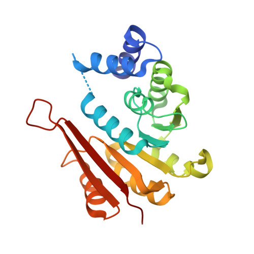
Crystal structure of mouse comt bound to an inhibitor


Experimental Data Snapshot

  • Method: X-RAY DIFFRACTION
  • Resolution: 2.06 Å
  • R-Value Free: 
    0.199 (Depositor), 0.242 (DCC) 
  • R-Value Work: 
    0.145 (Depositor) 
  • R-Value Observed: 
    0.147 (Depositor) 

Starting Model: experimental
View more details

wwPDB Validation 3D Report Full Report

Validation slider image for 4P58

Ligand Structure Quality Assessment 


This is version 1.2 of the entry. See complete history. 

Literature

A fragment-based approach to identifying S-adenosyl-l-methionine -competitive inhibitors of catechol O-methyl transferase (COMT).

Lanier, M., Ambrus, G., Cole, D.C., Davenport, R., Ellery, J., Fosbeary, R., Jennings, A.J., Kadotani, A., Kamada, Y., Kamran, R., Matsumoto, S., Mizukami, A., Okubo, S., Okada, K., Saikatendu, K., Walsh, L., Wu, H., Hixon, M.S.

(2014) J Med Chem 57: 5459-5463

  • DOI: https://doi.org/10.1021/jm500475k
  • Primary Citation Related Structures: 
    4P58

  • PubMed Abstract: 

    Catechol O-methyl transferase belongs to the diverse family of S-adenosyl-l-methionine transferases. It is a target involved in the treatment of Parkinson's disease. Here we present a fragment-based screening approach to discover noncatechol derived COMT inhibitors which bind at the SAM binding pocket. We describe the identification and characterization of a series of highly ligand efficient SAM competitive bisaryl fragments (LE = 0.33-0.58). We also present the first SAM-competitive small-molecule COMT co-complex crystal structure.


  • Organizational Affiliation: 
    • Medicinal Chemistry, ‡Structural Biology, §Discovery Biology, ∥Analytical Chemistry, Takeda California Inc. , 10410 Science Center Drive San Diego California 92121, United States.

Macromolecule Content 

  • Total Structure Weight: 24 kDa 
  • Atom Count: 1,784 
  • Modeled Residue Count: 205 
  • Deposited Residue Count: 212 
  • Unique protein chains: 1

Macromolecules

Find similar proteins by:|  3D Structure
Entity ID: 1
MoleculeChains  Sequence LengthOrganismDetailsImage
Catechol O-methyltransferase212Mus musculusMutation(s): 0 
Gene Names: Comt, Comt1
EC: 2.1.1.6
UniProt & NIH Common Fund Data Resources
Find proteins for O88587 (Mus musculus)
Explore O88587 
Go to UniProtKB:  O88587
IMPC:  MGI:88470
Entity Groups
Sequence Clusters30% Identity50% Identity70% Identity90% Identity95% Identity100% Identity
UniProt GroupO88587
Sequence Annotations
Expand
Reference Sequence

Small Molecules

Ligands 1 Unique
IDChains Name / Formula / InChI Key2D Diagram3D Interactions
2F6

Query on 2F6


Download:Ideal Coordinates CCD File
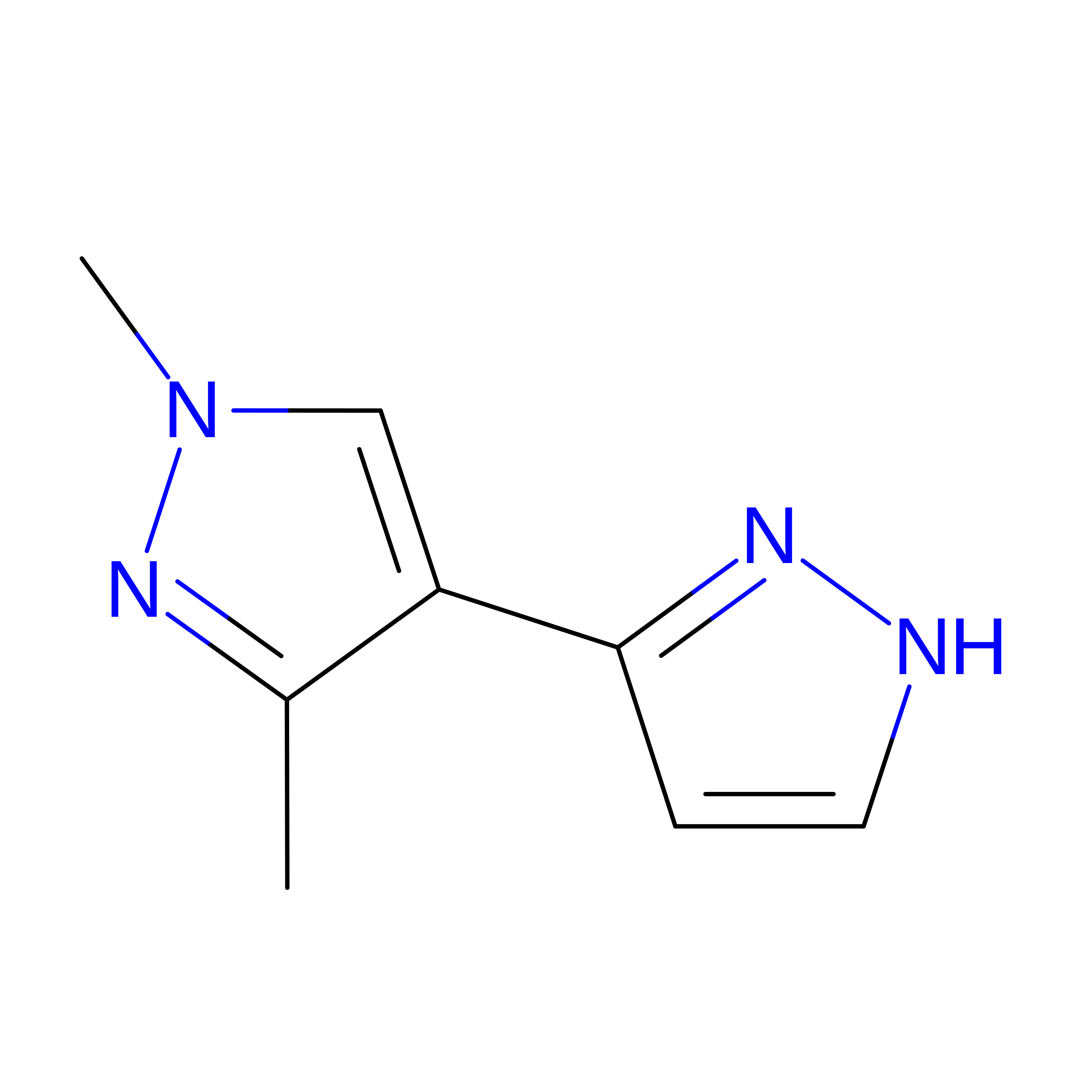
B [auth A]1',3'-dimethyl-1H,1'H-3,4'-bipyrazole
C8 H10 N4
LNJKDWJFZASOPC-UHFFFAOYSA-N

Experimental Data & Validation

Experimental Data

  • Method: X-RAY DIFFRACTION
  • Resolution: 2.06 Å
  • R-Value Free:  0.199 (Depositor), 0.242 (DCC) 
  • R-Value Work:  0.145 (Depositor) 
  • R-Value Observed: 0.147 (Depositor) 
Space Group: I 41 2 2
Unit Cell:
Length ( Å )Angle ( ˚ )
a = 98.584α = 90
b = 98.584β = 90
c = 115.104γ = 90
Software Package:
Software NamePurpose
REFMACrefinement
PDB_EXTRACTdata extraction

Structure Validation

View Full Validation Report


Ligand Structure Quality Assessment 


Entry History 

Deposition Data

  • Released Date: 2014-06-25 
  • Deposition Author(s): Lanier, M.

Revision History  (Full details and data files)

  • Version 1.0: 2014-06-25
    Type: Initial release
  • Version 1.1: 2014-10-01
    Changes: Database references
  • Version 1.2: 2023-09-27
    Changes: Data collection, Database references, Derived calculations, Other, Refinement description, Source and taxonomy, Structure summary