


Crystal Structure of Mycobacterium tuberculosis Shikimate Dehydrogenase
Lalgondar, M., Sacchettini, J.C.To be published.
Experimental Data Snapshot
Entity ID: 1 | |||||
|---|---|---|---|---|---|
| Molecule | Chains | Sequence Length | Organism | Details | Image |
| Shikimate 5-dehydrogenase AroE (5-dehydroshikimate reductase) | 277 | Mycobacterium tuberculosis | Mutation(s): 0 Gene Names: aroE, MT2629, Rv2552c EC: 1.1.1.25 | 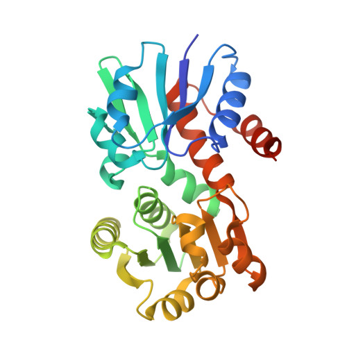 | |
UniProt | |||||
Entity Groups | |||||
| Sequence Clusters | 30% Identity50% Identity70% Identity90% Identity95% Identity100% Identity | ||||
| UniProt Group | I6Y120 | ||||
Sequence AnnotationsExpand | |||||
Reference Sequence | |||||
| Ligands 3 Unique | |||||
|---|---|---|---|---|---|
| ID | Chains | Name / Formula / InChI Key | 2D Diagram | 3D Interactions | |
| SKM Download:Ideal Coordinates CCD File | C [auth A] | (3R,4S,5R)-3,4,5-TRIHYDROXYCYCLOHEX-1-ENE-1-CARBOXYLIC ACID C7 H10 O5 JXOHGGNKMLTUBP-HSUXUTPPSA-N |  | ||
| SO4 Download:Ideal Coordinates CCD File | J [auth A], K [auth A], M [auth B] | SULFATE ION O4 S QAOWNCQODCNURD-UHFFFAOYSA-L |  | ||
| BR Download:Ideal Coordinates CCD File | D [auth A] E [auth A] F [auth A] G [auth A] H [auth A] | BROMIDE ION Br CPELXLSAUQHCOX-UHFFFAOYSA-M |  | ||
| Length ( Å ) | Angle ( ˚ ) |
|---|---|
| a = 43.467 | α = 90 |
| b = 75.553 | β = 90 |
| c = 129.708 | γ = 90 |
| Software Name | Purpose |
|---|---|
| PDB_EXTRACT | data extraction |
| PHENIX | refinement |
| Funding Organization | Location | Grant Number |
|---|---|---|
| National Institutes of Health/National Institute of General Medical Sciences (NIH/NIGMS) | United States | -- |


