4P4C | pdb_00004p4c

Human EphA3 Kinase domain in complex with quinoxaline derivatives


Experimental Data Snapshot

  • Method: X-RAY DIFFRACTION
  • Resolution: 1.60 Å
  • R-Value Free: 
    0.202 (Depositor), 0.202 (DCC) 
  • R-Value Work: 
    0.178 (Depositor), 0.179 (DCC) 
  • R-Value Observed: 
    0.179 (Depositor) 

Starting Model: experimental
View more details

wwPDB Validation 3D Report Full Report

Validation slider image for 4P4C

Ligand Structure Quality Assessment 


This is version 1.3 of the entry. See complete history

Literature

Pyrrolo[3,2-b]quinoxaline Derivatives as Types I1/2 and II Eph Tyrosine Kinase Inhibitors: Structure-Based Design, Synthesis, and in Vivo Validation.

Unzue, A.Dong, J.Lafleur, K.Zhao, H.Frugier, E.Caflisch, A.Nevado, C.

(2014) J Med Chem 57: 6834-6844

  • DOI: https://doi.org/10.1021/jm5009242
  • Primary Citation Related Structures: 
    4P4C, 4P5Q, 4P5Z

  • PubMed Abstract: 

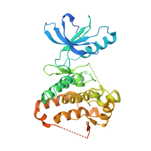
    The X-ray crystal structures of the catalytic domain of the EphA3 tyrosine kinase in complex with two type I inhibitors previously discovered in silico (compounds A and B) were used to design type I1/2 and II inhibitors. Chemical synthesis of about 25 derivatives culminated in the discovery of compounds 11d (type I1/2), 7b, and 7g (both of type II), which have low-nanomolar affinity for Eph kinases in vitro and a good selectivity profile on a panel of 453 human kinases (395 nonmutant). Surface plasmon resonance measurements show a very slow unbinding rate (1/115 min) for inhibitor 7m. Slow dissociation is consistent with a type II binding mode in which the hydrophobic moiety (trifluoromethyl-benzene) of the inhibitor is deeply buried in a cavity originating from the displacement of the Phe side chain of the so-called DFG motif as observed in the crystal structure of compound 7m. The inhibitor 11d displayed good in vivo efficacy in a human breast cancer xenograft.


  • Organizational Affiliation
    • Department of Chemistry and ‡Department of Biochemistry, University of Zürich , Winterthurerstrasse 190, CH-8057 Zürich, Switzerland.

Macromolecule Content 

  • Total Structure Weight: 40.79 kDa 
  • Atom Count: 2,409 
  • Modeled Residue Count: 266 
  • Deposited Residue Count: 361 
  • Unique protein chains: 1

Macromolecules

Find similar proteins by:|  3D Structure
Entity ID: 1
MoleculeChains  Sequence LengthOrganismDetailsImage
EPH receptor A3361Homo sapiensMutation(s): 0 
Gene Names: EPHA3
EC: 2.7.10.1
UniProt & NIH Common Fund Data Resources
Find proteins for P29320 (Homo sapiens)
Explore P29320 
Go to UniProtKB:  P29320
PHAROS:  P29320
GTEx:  ENSG00000044524 
Entity Groups
Sequence Clusters30% Identity50% Identity70% Identity90% Identity95% Identity100% Identity
UniProt GroupP29320
Sequence Annotations
Expand
Reference Sequence

Small Molecules

Ligands 1 Unique
IDChains Name / Formula / InChI Key2D Diagram3D Interactions
25Q

Query on 25Q


Download:Ideal Coordinates CCD File
B [auth A]2-amino-1-(3-methoxyphenyl)-1H-pyrrolo[2,3-b]quinoxaline-3-carboxamide
C18 H15 N5 O2
CHHKMVNVTYIPGZ-UHFFFAOYSA-N

Experimental Data & Validation

Experimental Data

  • Method: X-RAY DIFFRACTION
  • Resolution: 1.60 Å
  • R-Value Free:  0.202 (Depositor), 0.202 (DCC) 
  • R-Value Work:  0.178 (Depositor), 0.179 (DCC) 
  • R-Value Observed: 0.179 (Depositor) 
Space Group: P 1 21 1
Unit Cell:
Length ( Å )Angle ( ˚ )
a = 53.914α = 90
b = 38.164β = 101.59
c = 75.527γ = 90
Software Package:
Software NamePurpose
SCALAdata scaling
XDSdata scaling
PDB_EXTRACTdata extraction
PHASERphasing
PHENIXrefinement

Structure Validation

View Full Validation Report


Ligand Structure Quality Assessment 


Entry History 

& Funding Information

Deposition Data


Funding OrganizationLocationGrant Number
KrebsligaSwitzerland--
Swiss National Science FoundationSwitzerland--

Revision History  (Full details and data files)

  • Version 1.0: 2014-08-13
    Type: Initial release
  • Version 1.1: 2014-10-01
    Changes: Database references
  • Version 1.2: 2017-11-22
    Changes: Database references, Derived calculations, Other, Refinement description, Source and taxonomy
  • Version 1.3: 2023-09-27
    Changes: Data collection, Database references, Refinement description