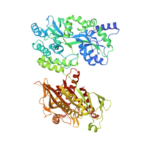
4OZQ | pdb_00004ozq

Crystal structure of the mouse Kif14 motor domain


Experimental Data Snapshot

  • Method: X-RAY DIFFRACTION
  • Resolution: 2.71 Å
  • R-Value Free: 
    0.307 (Depositor), 0.290 (DCC) 
  • R-Value Work: 
    0.264 (Depositor), 0.240 (DCC) 
  • R-Value Observed: 
    0.266 (Depositor) 

wwPDB Validation   3D Report Full Report


Ligand Structure Quality Assessment 

Created with Raphaël 2.3.0Worse 01 BetterLigand structure goodness of fit to experimental dataBest fitted ADPClick on this verticalbar to view details

This is version 1.3 of the entry. See complete history


Literature

KIF14 binds tightly to microtubules and adopts a rigor-like conformation.

Arora, K.Talje, L.Asenjo, A.B.Andersen, P.Atchia, K.Joshi, M.Sosa, H.Allingham, J.S.Kwok, B.H.

(2014) J Mol Biology 426: 2997-3015

  • DOI: https://doi.org/10.1016/j.jmb.2014.05.030
  • Primary Citation of Related Structures:  
    4OZQ

  • PubMed Abstract: 

    The mitotic kinesin motor protein KIF14 is essential for cytokinesis during cell division and has been implicated in cerebral development and a variety of human cancers. Here we show that the mouse KIF14 motor domain binds tightly to microtubules and does not display typical nucleotide-dependent changes in this affinity. It also has robust ATPase activity but very slow motility. A crystal structure of the ADP-bound form of the KIF14 motor domain reveals a dramatically opened ATP-binding pocket, as if ready to exchange its bound ADP for Mg·ATP. In this state, the central β-sheet is twisted ~10° beyond the maximal amount observed in other kinesins. This configuration has only been seen in the nucleotide-free states of myosins-known as the "rigor-like" state. Fitting of this atomic model to electron density maps from cryo-electron microscopy indicates a distinct binding configuration of the motor domain to microtubules. We postulate that these properties of KIF14 are well suited for stabilizing midbody microtubules during cytokinesis.


  • Organizational Affiliation

    Department of Biomedical and Molecular Sciences, Queen's University, 18 Stuart St., Rm. 652, Kingston, ON K7L 3 N6, Canada.


Macromolecules
Find similar proteins by:  (by identity cutoff)  |  3D Structure
Entity ID: 1
MoleculeChains Sequence LengthOrganismDetailsImage
Chimera of Maltose-binding periplasmic protein and Kinesin family member 14 protein
A, B
720Escherichia coli UMEA 3304-1Mus musculus
This entity is chimeric
Mutation(s): 0 
Gene Names: G962_03763Kif14
UniProt
Find proteins for L0N7N1 (Mus musculus)
Explore L0N7N1 
Go to UniProtKB:  L0N7N1
Entity Groups  
Sequence Clusters30% Identity50% Identity70% Identity90% Identity95% Identity100% Identity
UniProt GroupL0N7N1
Sequence Annotations
Expand
  • Reference Sequence
Experimental Data & Validation

Experimental Data

  • Method: X-RAY DIFFRACTION
  • Resolution: 2.71 Å
  • R-Value Free:  0.307 (Depositor), 0.290 (DCC) 
  • R-Value Work:  0.264 (Depositor), 0.240 (DCC) 
  • R-Value Observed: 0.266 (Depositor) 
Space Group: P 1
Unit Cell:
Length ( Å )Angle ( ˚ )
a = 65.137α = 74.43
b = 72.193β = 87.45
c = 94.098γ = 89.94
Software Package:
Software NamePurpose
REFMACrefinement

Structure Validation

View Full Validation Report


Ligand Structure Quality Assessment 

Created with Raphaël 2.3.0Worse 01 BetterLigand structure goodness of fit to experimental dataBest fitted ADPClick on this verticalbar to view details

Entry History & Funding Information

Deposition Data


Funding OrganizationLocationGrant Number
Canadian Institutes of HealthCanada--

Revision History  (Full details and data files)

  • Version 1.0: 2014-07-09
    Type: Initial release
  • Version 1.1: 2014-10-01
    Changes: Database references
  • Version 1.2: 2015-02-04
    Changes: Derived calculations
  • Version 1.3: 2023-12-27
    Changes: Advisory, Data collection, Database references, Derived calculations, Other, Refinement description, Source and taxonomy