4OUL | pdb_00004oul

Crystal structure of human Caprin-2 C1q domain


Experimental Data Snapshot

  • Method: X-RAY DIFFRACTION
  • Resolution: 1.95 Å
  • R-Value Free: 
    0.251 (Depositor), 0.252 (DCC) 
  • R-Value Work: 
    0.203 (Depositor), 0.208 (DCC) 
  • R-Value Observed: 
    0.205 (Depositor) 

wwPDB Validation 3D Report Full Report

Validation slider image for 4OUL

This is version 1.2 of the entry. See complete history

Literature

Structural insights into the C1q domain of Caprin-2 in canonical Wnt signaling

Miao, H.Jia, Y.Xie, S.Wang, X.Zhao, J.Chu, Y.Zhou, Z.Shi, Z.Song, X.Li, L.

(2014) J Biological Chem 289: 34104-34113

  • DOI: https://doi.org/10.1074/jbc.M114.591636
  • Primary Citation Related Structures: 
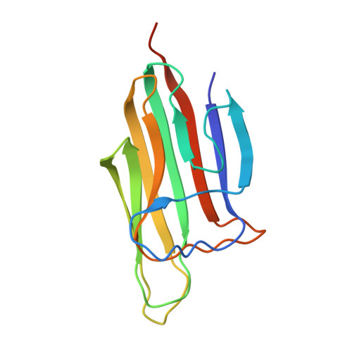
    4OUL, 4OUM, 4OUS

  • PubMed Abstract: 

    Previously, we have identified Caprin-2 as a new regulator in canonical Wnt signaling through a mechanism of facilitating LRP5/6 phosphorylation; moreover, we found that its C-terminal C1q-related domain (Cap2_CRD) is required for this process. Here, we determined the crystal structures of Cap2_CRD from human and zebrafish, which both associate as a homotrimer with calcium located at the symmetric center. Surprisingly, the calcium binding-deficient mutant exists as a more stable trimer than its wild-type counterpart. Further studies showed that this Caprin-2 mutant disabled in binding calcium maintains the activity of promoting LRP5/6 phosphorylation, whereas the mutations disrupting Cap2_CRD homotrimer did impair such activity. Together, our findings suggested that the C-terminal CRD domain of Caprin-2 forms a flexible homotrimer mediated by calcium and that such trimeric assembly is required for Caprin-2 to regulate canonical Wnt signaling.


  • Organizational Affiliation
    • From the State Key Laboratory of Molecular Biology, Institute of Biochemistry and Cell Biology, Shanghai Institutes for Biological Sciences, Chinese Academy of Sciences, Shanghai 200031, China.

Macromolecule Content 

  • Total Structure Weight: 95.82 kDa 
  • Atom Count: 6,291 
  • Modeled Residue Count: 789 
  • Deposited Residue Count: 840 
  • Unique protein chains: 1

Macromolecules

Find similar proteins by:|  3D Structure
Entity ID: 1
MoleculeChains  Sequence LengthOrganismDetailsImage
Caprin-2
A, B, C, D, E
A, B, C, D, E, F
140Homo sapiensMutation(s): 0 
Gene Names: C1QDC1CAPRIN2EEG1KIAA1873RNG140
UniProt & NIH Common Fund Data Resources
Find proteins for Q6IMN6 (Homo sapiens)
Explore Q6IMN6 
Go to UniProtKB:  Q6IMN6
PHAROS:  Q6IMN6
GTEx:  ENSG00000110888 
Entity Groups
Sequence Clusters30% Identity50% Identity70% Identity90% Identity95% Identity100% Identity
UniProt GroupQ6IMN6
Sequence Annotations
Expand
Reference Sequence

Experimental Data & Validation

Experimental Data

  • Method: X-RAY DIFFRACTION
  • Resolution: 1.95 Å
  • R-Value Free:  0.251 (Depositor), 0.252 (DCC) 
  • R-Value Work:  0.203 (Depositor), 0.208 (DCC) 
  • R-Value Observed: 0.205 (Depositor) 
Space Group: P 1
Unit Cell:
Length ( Å )Angle ( ˚ )
a = 48.353α = 89.51
b = 48.342β = 87.24
c = 80.884γ = 71.74
Software Package:
Software NamePurpose
HKL-2000data collection
PHASERphasing
PHENIXrefinement
HKL-2000data reduction
HKL-2000data scaling

Structure Validation

View Full Validation Report


Entry History 

Deposition Data

Revision History  (Full details and data files)

  • Version 1.0: 2014-10-29
    Type: Initial release
  • Version 1.1: 2015-04-08
    Changes: Database references
  • Version 1.2: 2024-03-20
    Changes: Data collection, Database references, Derived calculations