4OSO

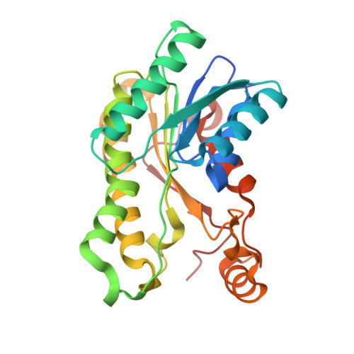
The crystal structure of landomycin C-6 ketoreductase LanV with bound NADP and rabelomycin


Experimental Data Snapshot

  • Method: X-RAY DIFFRACTION
  • Resolution: 2.50 Å
  • R-Value Free: 0.235 
  • R-Value Work: 0.174 
  • R-Value Observed: 0.178 

Starting Model: experimental
View more details

wwPDB Validation   3D Report Full Report


Ligand Structure Quality Assessment 


This is version 1.3 of the entry. See complete history


Literature

Structure-based engineering of angucyclinone 6-ketoreductases.

Patrikainen, P.Niiranen, L.Thapa, K.Paananen, P.Tahtinen, P.Mantsala, P.Niemi, J.Metsa-Ketela, M.

(2014) Chem Biol 21: 1381-1391

  • DOI: https://doi.org/10.1016/j.chembiol.2014.07.017
  • Primary Citation of Related Structures:  
    4OSO, 4OSP

  • PubMed Abstract: 

    Angucyclines are tetracyclic polyketides produced by Streptomyces bacteria that exhibit notable biological activities. The great diversity of angucyclinones is generated in tailoring reactions, which modify the common benz[a]anthraquinone carbon skeleton. In particular, the opposite stereochemistry of landomycins and urdamycins/gaudimycins at C-6 is generated by the short-chain alcohol dehydrogenases/reductases LanV and UrdMred/CabV, respectively. Here we present crystal structures of LanV and UrdMred in complex with NADP(+) and the product analog rabelomycin, which enabled us to identify four regions associated with the functional differentiation. The structural analysis was confirmed in chimeragenesis experiments focusing on these regions adjacent to the active site cavity, which led to reversal of the activities of LanV and CabV. The results surprisingly indicated that the conformation of the substrate and the stereochemical outcome of 6-ketoreduction appear to be intimately linked.


  • Organizational Affiliation

    Department of Biochemistry, University of Turku, 20014 Turku, Finland.


Macromolecules
Find similar proteins by:  (by identity cutoff)  |  3D Structure
Entity ID: 1
MoleculeChains Sequence LengthOrganismDetailsImage
Reductase homolog
A, B
263Streptomyces cyanogenusMutation(s): 0 
Gene Names: lanV
UniProt
Find proteins for Q9ZGC1 (Streptomyces cyanogenus)
Explore Q9ZGC1 
Go to UniProtKB:  Q9ZGC1
Entity Groups  
Sequence Clusters30% Identity50% Identity70% Identity90% Identity95% Identity100% Identity
UniProt GroupQ9ZGC1
Sequence Annotations
Expand
  • Reference Sequence
Small Molecules
Ligands 3 Unique
IDChains Name / Formula / InChI Key2D Diagram3D Interactions
NAP
Query on NAP

Download Ideal Coordinates CCD File 
C [auth A],
F [auth B]
NADP NICOTINAMIDE-ADENINE-DINUCLEOTIDE PHOSPHATE
C21 H28 N7 O17 P3
XJLXINKUBYWONI-NNYOXOHSSA-N
2V4
Query on 2V4

Download Ideal Coordinates CCD File 
D [auth A],
G [auth B]
rabelomycin
C19 H14 O6
JJOLHRYZQSDLSA-LJQANCHMSA-N
PEG
Query on PEG

Download Ideal Coordinates CCD File 
E [auth A],
H [auth B]
DI(HYDROXYETHYL)ETHER
C4 H10 O3
MTHSVFCYNBDYFN-UHFFFAOYSA-N
Experimental Data & Validation

Experimental Data

  • Method: X-RAY DIFFRACTION
  • Resolution: 2.50 Å
  • R-Value Free: 0.235 
  • R-Value Work: 0.174 
  • R-Value Observed: 0.178 
  • Space Group: P 41 21 2
Unit Cell:
Length ( Å )Angle ( ˚ )
a = 91.99α = 90
b = 91.99β = 90
c = 106.22γ = 90
Software Package:
Software NamePurpose
XSCALEdata scaling
MOLREPphasing
REFMACrefinement
PDB_EXTRACTdata extraction
EDNAdata collection
XDSdata reduction

Structure Validation

View Full Validation Report


Ligand Structure Quality Assessment 


Entry History 

Deposition Data

Revision History  (Full details and data files)

  • Version 1.0: 2014-10-01
    Type: Initial release
  • Version 1.1: 2014-11-12
    Changes: Structure summary
  • Version 1.2: 2014-12-03
    Changes: Database references
  • Version 1.3: 2023-09-20
    Changes: Data collection, Database references, Derived calculations, Refinement description