4OR8 | pdb_00004or8

Crystal structure of Marburg virus VP24


Experimental Data Snapshot

  • Method: X-RAY DIFFRACTION
  • Resolution: 2.65 Å
  • R-Value Free: 
    0.255 (Depositor), 0.261 (DCC) 
  • R-Value Work: 
    0.180 (Depositor), 0.187 (DCC) 
  • R-Value Observed: 
    0.183 (Depositor) 

wwPDB Validation 3D Report Full Report

Validation slider image for 4OR8

This is version 1.2 of the entry. See complete history

Literature

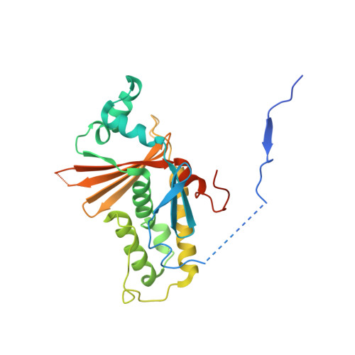
Crystal Structure of Marburg Virus VP24.

Zhang, A.P.Bornholdt, Z.A.Abelson, D.M.Saphire, E.O.

(2014) J Virol 88: 5859-5863

  • DOI: https://doi.org/10.1128/JVI.03565-13
  • Primary Citation Related Structures: 
    4OR8

  • PubMed Abstract: 

    The VP24 protein plays an essential, albeit poorly understood role in the filovirus life cycle. VP24 is only 30% identical between Marburg virus and the ebolaviruses. Furthermore, VP24 from the ebolaviruses is immunosuppressive, while that of Marburg virus is not. The crystal structure of Marburg virus VP24, presented here, reveals that although the core is similar between the viral genera, Marburg VP24 is distinguished by a projecting β-shelf and an alternate conformation of the N-terminal polypeptide.


  • Organizational Affiliation
    • Department of Immunology and Microbial Science, The Scripps Research Institute, La Jolla, California, USA.

Macromolecule Content 

  • Total Structure Weight: 60.77 kDa 
  • Atom Count: 3,682 
  • Modeled Residue Count: 451 
  • Deposited Residue Count: 534 
  • Unique protein chains: 1

Macromolecules

Find similar proteins by:|  3D Structure
Entity ID: 1
MoleculeChains  Sequence LengthOrganismDetailsImage
Membrane-associated protein VP24
A, B
267Marburg virus - Musoke, Kenya, 1980Mutation(s): 0 
Gene Names: VP24
UniProt
Find proteins for P35256 (Lake Victoria marburgvirus (strain Musoke-80))
Explore P35256 
Go to UniProtKB:  P35256
Entity Groups
Sequence Clusters30% Identity50% Identity70% Identity90% Identity95% Identity100% Identity
UniProt GroupP35256
Sequence Annotations
Expand
Reference Sequence

Experimental Data & Validation

Experimental Data

  • Method: X-RAY DIFFRACTION
  • Resolution: 2.65 Å
  • R-Value Free:  0.255 (Depositor), 0.261 (DCC) 
  • R-Value Work:  0.180 (Depositor), 0.187 (DCC) 
  • R-Value Observed: 0.183 (Depositor) 
Space Group: P 1
Unit Cell:
Length ( Å )Angle ( ˚ )
a = 42.221α = 88.44
b = 48.078β = 78.68
c = 64.051γ = 71.34
Software Package:
Software NamePurpose
HKL-2000data collection
PHASERphasing
PHENIXrefinement
HKL-2000data reduction
HKL-2000data scaling

Structure Validation

View Full Validation Report


Entry History 

Deposition Data

Revision History  (Full details and data files)

  • Version 1.0: 2014-03-26
    Type: Initial release
  • Version 1.1: 2014-05-07
    Changes: Database references
  • Version 1.2: 2024-02-28
    Changes: Data collection, Database references