4OR2 | pdb_00004or2

Human class C G protein-coupled metabotropic glutamate receptor 1 in complex with a negative allosteric modulator


Experimental Data Snapshot

  • Method: X-RAY DIFFRACTION
  • Resolution: 2.80 Å
  • R-Value Free: 
    0.268 (Depositor), 0.273 (DCC) 
  • R-Value Work: 
    0.227 (Depositor) 
  • R-Value Observed: 
    0.229 (Depositor) 

Starting Model: experimental
View more details

wwPDB Validation 3D Report Full Report

Validation slider image for 4OR2

Ligand Structure Quality Assessment 


This is version 1.7 of the entry. See complete history

Literature

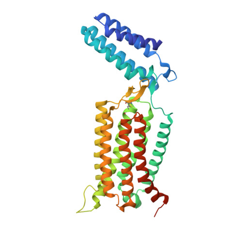
Structure of a class C GPCR metabotropic glutamate receptor 1 bound to an allosteric modulator

Wu, H.Wang, C.Gregory, K.J.Han, G.W.Cho, H.P.Xia, Y.Niswender, C.M.Katritch, V.Meiler, J.Cherezov, V.Conn, P.J.Stevens, R.C.

(2014) Science 344: 58-64

  • DOI: https://doi.org/10.1126/science.1249489
  • Primary Citation Related Structures: 
    4OR2

  • PubMed Abstract: 

    The excitatory neurotransmitter glutamate induces modulatory actions via the metabotropic glutamate receptors (mGlus), which are class C G protein-coupled receptors (GPCRs). We determined the structure of the human mGlu1 receptor seven-transmembrane (7TM) domain bound to a negative allosteric modulator, FITM, at a resolution of 2.8 angstroms. The modulator binding site partially overlaps with the orthosteric binding sites of class A GPCRs but is more restricted than most other GPCRs. We observed a parallel 7TM dimer mediated by cholesterols, which suggests that signaling initiated by glutamate's interaction with the extracellular domain might be mediated via 7TM interactions within the full-length receptor dimer. A combination of crystallography, structure-activity relationships, mutagenesis, and full-length dimer modeling provides insights about the allosteric modulation and activation mechanism of class C GPCRs.


  • Organizational Affiliation
    • Department of Integrative Structural and Computational Biology, The Scripps Research Institute, 10550 North Torrey Pines Road, La Jolla, CA 92037, USA.

Macromolecule Content 

  • Total Structure Weight: 91.11 kDa 
  • Atom Count: 5,906 
  • Modeled Residue Count: 726 
  • Deposited Residue Count: 778 
  • Unique protein chains: 1

Macromolecules

Find similar proteins by:|  3D Structure
Entity ID: 1
MoleculeChains  Sequence LengthOrganismDetailsImage
Soluble cytochrome b562, Metabotropic glutamate receptor 1
A, B
389Escherichia coliHomo sapiens
This entity is chimeric
Mutation(s): 3 
Gene Names: cybCGRM1_HUMANGPRC1AGRM1MGLUR1
Membrane Entity: Yes 
UniProt & NIH Common Fund Data Resources
Find proteins for P0ABE7 (Escherichia coli)
Explore P0ABE7 
Go to UniProtKB:  P0ABE7
Find proteins for Q13255 (Homo sapiens)
Explore Q13255 
Go to UniProtKB:  Q13255
PHAROS:  Q13255
GTEx:  ENSG00000152822 
Entity Groups
Sequence Clusters30% Identity50% Identity70% Identity90% Identity95% Identity100% Identity
UniProt GroupsP0ABE7Q13255
Sequence Annotations
Expand
Reference Sequence

Small Molecules

Ligands 5 Unique
IDChains Name / Formula / InChI Key2D Diagram3D Interactions
CLR

Query on CLR


Download:Ideal Coordinates CCD File
D [auth A]
E [auth A]
F [auth A]
G [auth A]
O [auth B]
D [auth A],
E [auth A],
F [auth A],
G [auth A],
O [auth B],
P [auth B]
CHOLESTEROL
C27 H46 O
HVYWMOMLDIMFJA-DPAQBDIFSA-N
FM9

Query on FM9


Download:Ideal Coordinates CCD File
C [auth A],
K [auth B]
4-fluoro-N-methyl-N-{4-[6-(propan-2-ylamino)pyrimidin-4-yl]-1,3-thiazol-2-yl}benzamide
C18 H18 F N5 O S
WIVGIKIKQHUFOD-UHFFFAOYSA-N
OLC

Query on OLC


Download:Ideal Coordinates CCD File
I [auth A],
J [auth A],
N [auth B]
(2R)-2,3-dihydroxypropyl (9Z)-octadec-9-enoate
C21 H40 O4
RZRNAYUHWVFMIP-GDCKJWNLSA-N
OLA

Query on OLA


Download:Ideal Coordinates CCD File
H [auth A]OLEIC ACID
C18 H34 O2
ZQPPMHVWECSIRJ-KTKRTIGZSA-N
PO4

Query on PO4


Download:Ideal Coordinates CCD File
L [auth B],
M [auth B]
PHOSPHATE ION
O4 P
NBIIXXVUZAFLBC-UHFFFAOYSA-K

Experimental Data & Validation

Experimental Data

  • Method: X-RAY DIFFRACTION
  • Resolution: 2.80 Å
  • R-Value Free:  0.268 (Depositor), 0.273 (DCC) 
  • R-Value Work:  0.227 (Depositor) 
  • R-Value Observed: 0.229 (Depositor) 
Space Group: P 21 21 21
Unit Cell:
Length ( Å )Angle ( ˚ )
a = 67.361α = 90
b = 86.552β = 90
c = 168.277γ = 90
Software Package:
Software NamePurpose
Blu-Icedata collection
AutoSolphasing
BUSTERrefinement
HKL-2000data reduction
HKL-2000data scaling

Structure Validation

View Full Validation Report


Ligand Structure Quality Assessment 


Entry History 

Revision History  (Full details and data files)

  • Version 1.0: 2014-03-19
    Type: Initial release
  • Version 1.1: 2014-03-26
    Changes: Structure summary
  • Version 1.2: 2015-02-04
    Changes: Derived calculations
  • Version 1.3: 2015-04-01
    Changes: Database references
  • Version 1.4: 2017-08-16
    Changes: Refinement description, Source and taxonomy
  • Version 1.5: 2017-11-22
    Changes: Refinement description
  • Version 1.6: 2024-04-03
    Changes: Data collection, Database references, Derived calculations, Refinement description
  • Version 1.7: 2024-11-13
    Changes: Structure summary