


Crystal structure of a hypothetical leucine rich repeat protein (BACCAP_00569) from Bacteroides capillosus ATCC 29799 at 2.00 A resolution
Joint Center for Structural Genomics (JCSG)To be published.
Experimental Data Snapshot
wwPDB Validation 3D Report Full Report

Entity ID: 1 | |||||
|---|---|---|---|---|---|
| Molecule | Chains | Sequence Length | Organism | Details | Image |
| hypothetical leucine rich repeat protein | 148 | Pseudoflavonifractor capillosus ATCC 29799 | Mutation(s): 0 Gene Names: BACCAP_00569, ZP_02034978.1 | 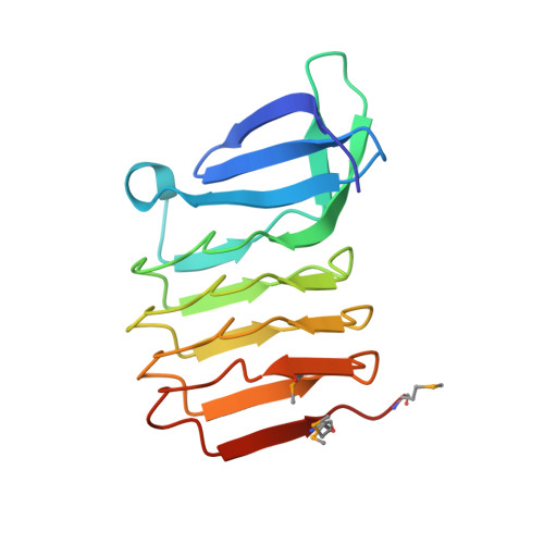 | |
UniProt | |||||
Entity Groups | |||||
| Sequence Clusters | 30% Identity50% Identity70% Identity90% Identity95% Identity100% Identity | ||||
| UniProt Group | A6NQU6 | ||||
Sequence AnnotationsExpand | |||||
Reference Sequence | |||||
| Ligands 2 Unique | |||||
|---|---|---|---|---|---|
| ID | Chains | Name / Formula / InChI Key | 2D Diagram | 3D Interactions | |
| EDO Download:Ideal Coordinates CCD File | E [auth A] F [auth A] G [auth A] J [auth B] K [auth B] | 1,2-ETHANEDIOL C2 H6 O2 LYCAIKOWRPUZTN-UHFFFAOYSA-N |  | ||
| MG Download:Ideal Coordinates CCD File | H [auth A], I [auth A], N [auth C] | MAGNESIUM ION Mg JLVVSXFLKOJNIY-UHFFFAOYSA-N |  | ||
| Modified Residues 1 Unique | |||||
|---|---|---|---|---|---|
| ID | Chains | Type | Formula | 2D Diagram | Parent |
| MSE Query on MSE | A, B, C, D | L-PEPTIDE LINKING | C5 H11 N O2 Se |  | MET |
| Length ( Å ) | Angle ( ˚ ) |
|---|---|
| a = 57.635 | α = 90 |
| b = 108.16 | β = 90 |
| c = 108.192 | γ = 90 |
| Software Name | Purpose |
|---|---|
| MolProbity | model building |
| PDB_EXTRACT | data extraction |
| SHELX | phasing |
| SHARP | phasing |
| XSCALE | data scaling |
| BUSTER-TNT | refinement |
| XDS | data reduction |
| SHELXD | phasing |
| BUSTER | refinement |


