4OFB | pdb_00004ofb

Crystal structure of human BRCA1 BRCT in complex with nonphosphopeptide inhibitor


Experimental Data Snapshot

  • Method: X-RAY DIFFRACTION
  • Resolution: 3.05 Å
  • R-Value Free: 
    0.237 (Depositor), 0.237 (DCC) 
  • R-Value Work: 
    0.210 (Depositor), 0.213 (DCC) 
  • R-Value Observed: 
    0.212 (Depositor) 

wwPDB Validation 3D Report Full Report

Validation slider image for 4OFB

This is version 1.3 of the entry. See complete history

Literature

Peptide Library Approach to Uncover Phosphomimetic Inhibitors of the BRCA1 C-Terminal Domain.

White, E.R.Sun, L.Ma, Z.Beckta, J.M.Danzig, B.A.Hacker, D.E.Huie, M.Williams, D.C.Edwards, R.A.Valerie, K.Glover, J.N.Hartman, M.C.

(2015) ACS Chem Biol 10: 1198-1208

  • DOI: https://doi.org/10.1021/cb500757u
  • Primary Citation Related Structures: 
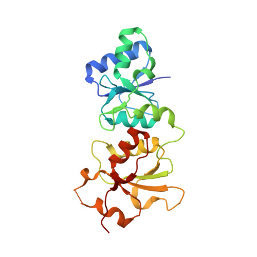
    4OFB

  • PubMed Abstract: 

    Many intracellular protein-protein interactions are mediated by the phosphorylation of serine, and phosphoserine-containing peptides can inhibit these interactions. However, hydrolysis of the phosphate by phosphatases, and the poor cell permeability associated with phosphorylated peptides has limited their utility in cellular and in vivo contexts. Compounding the problem, strategies to replace phosphoserine in peptide inhibitors with easily accessible mimetics (such as Glu or Asp) routinely fail. Here, we present an in vitro selection strategy for replacement of phosphoserine. Using mRNA display, we created a 10 trillion member structurally diverse unnatural peptide library. From this library, we found a peptide that specifically binds to the C-terminal domain (BRCT)2 of breast cancer associated protein 1 (BRCA1) with an affinity comparable to phosphorylated peptides. A crystal structure of the peptide bound reveals that the pSer-x-x-Phe motif normally found in BRCA1 (BRCT)2 binding partners is replaced by a Glu-x-x-4-fluoroPhe and that the peptide picks up additional contacts on the protein surface not observed in cognate phosphopeptide binding. Expression of the peptide in human cells led to defects in DNA repair by homologous recombination, a process BRCA1 is known to coordinate. Overall, this work validates a new in vitro selection approach for the development of inhibitors of protein-protein interactions mediated by serine phosphorylation.


  • Organizational Affiliation
    • †Department of Chemistry, Virginia Commonwealth University (VCU), 1001 West Main Street, P.O. Box 842006, Richmond, Virginia 23284, United States.

Macromolecule Content 

  • Total Structure Weight: 26.19 kDa 
  • Atom Count: 1,791 
  • Modeled Residue Count: 223 
  • Deposited Residue Count: 228 
  • Unique protein chains: 2

Macromolecules

Find similar proteins by:|  3D Structure
Entity ID: 1
MoleculeChains  Sequence LengthOrganismDetailsImage
Breast cancer type 1 susceptibility protein214Homo sapiensMutation(s): 0 
Gene Names: BRCA1RNF53
EC: 2.3.2.27
UniProt & NIH Common Fund Data Resources
Find proteins for P38398 (Homo sapiens)
Explore P38398 
Go to UniProtKB:  P38398
PHAROS:  P38398
GTEx:  ENSG00000012048 
Entity Groups
Sequence Clusters30% Identity50% Identity70% Identity90% Identity95% Identity100% Identity
UniProt GroupP38398
Sequence Annotations
Expand
Reference Sequence
Find similar proteins by:  Sequence   |   3D Structure  
Entity ID: 2
MoleculeChains  Sequence LengthOrganismDetailsImage
nonphosphopeptide inhibitor14N/AMutation(s): 0 

Small Molecules

Modified Residues  1 Unique
IDChains TypeFormula2D DiagramParent
PFF
Query on PFF
B
L-PEPTIDE LINKINGC9 H10 F N O2PHE

Experimental Data & Validation

Experimental Data

  • Method: X-RAY DIFFRACTION
  • Resolution: 3.05 Å
  • R-Value Free:  0.237 (Depositor), 0.237 (DCC) 
  • R-Value Work:  0.210 (Depositor), 0.213 (DCC) 
  • R-Value Observed: 0.212 (Depositor) 
Space Group: P 41
Unit Cell:
Length ( Å )Angle ( ˚ )
a = 37.811α = 90
b = 37.811β = 90
c = 175.993γ = 90
Software Package:
Software NamePurpose
DENZOdata reduction
SCALEPACKdata scaling
PHASERphasing
PHENIXrefinement
PDB_EXTRACTdata extraction
MxDCdata collection

Structure Validation

View Full Validation Report


Entry History 

Deposition Data

Revision History  (Full details and data files)

  • Version 1.0: 2015-04-15
    Type: Initial release
  • Version 1.1: 2015-05-27
    Changes: Database references
  • Version 1.2: 2017-11-22
    Changes: Refinement description
  • Version 1.3: 2025-03-26
    Changes: Data collection, Database references, Derived calculations, Structure summary