4OC6 | pdb_00004oc6

Structure of Cathepsin D with inhibitor 2-bromo-N-[(2S,3S)-4-{[2-(2,4-dichlorophenyl)ethyl][3-(1,3-dioxo-1,3-dihydro-2H-isoindol-2-yl)propanoyl]amino}-3-hydroxy-1-(3-phenoxyphenyl)butan-2-yl]-4,5-dimethoxybenzamide


Experimental Data Snapshot

  • Method: X-RAY DIFFRACTION
  • Resolution: 2.64 Å
  • R-Value Free: 
    0.282 (Depositor), 0.288 (DCC) 
  • R-Value Work: 
    0.207 (Depositor), 0.206 (DCC) 
  • R-Value Observed: 
    0.211 (Depositor) 

wwPDB Validation 3D Report Full Report

Validation slider image for 4OC6

Ligand Structure Quality Assessment 


This is version 2.1 of the entry. See complete history

Literature

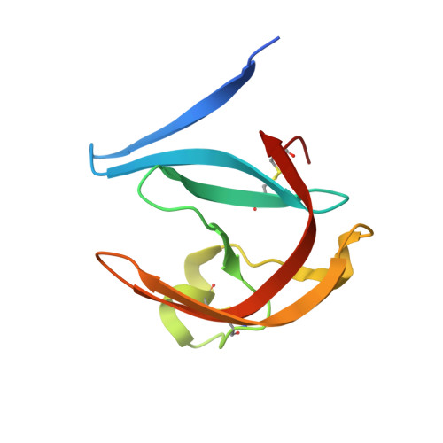
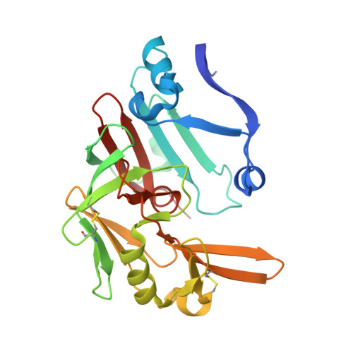
Structure-based optimization of non-peptidic Cathepsin D inhibitors.

Gradler, U.Czodrowski, P.Tsaklakidis, C.Klein, M.Werkmann, D.Lindemann, S.Maskos, K.Leuthner, B.

(2014) Bioorg Med Chem Lett 24: 4141-4150

  • DOI: https://doi.org/10.1016/j.bmcl.2014.07.054
  • Primary Citation Related Structures: 
    4OBZ, 4OC6, 4OD9

  • PubMed Abstract: 

    We discovered a novel series of non-peptidic acylguanidine inhibitors of Cathepsin D as target for osteoarthritis. The initial HTS-hits were optimized by structure-based design using CatD X-ray structures resulting in single digit nanomolar potency in the biochemical CatD assay. However, the most potent analogues showed only micromolar activities in an ex vivo glycosaminoglycan (GAG) release assay in bovine cartilage together with low cellular permeability and suboptimal microsomal stability. This new scaffold can serve as a starting point for further optimization towards in vivo efficacy.


  • Organizational Affiliation
    • Merck KGaA, Merck Serono Research, Small Molecule Platform, Frankfurter Str. 250, 64293 Darmstadt, Germany. Electronic address: ulrich.graedler@merckgroup.com.

Macromolecule Content 

  • Total Structure Weight: 39.77 kDa 
  • Atom Count: 2,758 
  • Modeled Residue Count: 341 
  • Deposited Residue Count: 346 
  • Unique protein chains: 2

Macromolecules

Find similar proteins by:|  3D Structure
Entity ID: 1
MoleculeChains  Sequence LengthOrganismDetailsImage
Cathepsin D light chain103Homo sapiensMutation(s): 0 
Gene Names: CTSDCPSD
EC: 3.4.23.5
UniProt & NIH Common Fund Data Resources
Find proteins for P07339 (Homo sapiens)
Explore P07339 
Go to UniProtKB:  P07339
PHAROS:  P07339
GTEx:  ENSG00000117984 
Entity Groups
Sequence Clusters30% Identity50% Identity70% Identity90% Identity95% Identity100% Identity
UniProt GroupP07339
Glycosylation
Glycosylation Sites: 1Go to GlyGen: P07339-1
Sequence Annotations
Expand
Reference Sequence
Find similar proteins by:|  3D Structure
Entity ID: 2
MoleculeChains  Sequence LengthOrganismDetailsImage
Cathepsin D heavy chain243Homo sapiensMutation(s): 0 
Gene Names: CTSDCPSD
EC: 3.4.23.5
UniProt & NIH Common Fund Data Resources
Find proteins for P07339 (Homo sapiens)
Explore P07339 
Go to UniProtKB:  P07339
PHAROS:  P07339
GTEx:  ENSG00000117984 
Entity Groups
Sequence Clusters30% Identity50% Identity70% Identity90% Identity95% Identity100% Identity
UniProt GroupP07339
Glycosylation
Glycosylation Sites: 1Go to GlyGen: P07339-1
Sequence Annotations
Expand
Reference Sequence

Oligosaccharides

Help  
Entity ID: 3
MoleculeChains Length2D Diagram GlycosylationD Interactions
alpha-D-mannopyranose-(1-6)-beta-D-mannopyranose-(1-4)-2-acetamido-2-deoxy-beta-D-glucopyranose-(1-4)-2-acetamido-2-deoxy-beta-D-glucopyranose
C
4N-Glycosylation
Glycosylation Resources
GlyTouCan: G22573RC
GlyCosmos: G22573RC
GlyGen: G22573RC

Small Molecules

Ligands 3 Unique
IDChains Name / Formula / InChI Key2D Diagram3D Interactions
2S1

Query on 2S1


Download:Ideal Coordinates CCD File
E [auth B]2-bromo-N-[(2S,3S)-4-{[2-(2,4-dichlorophenyl)ethyl][3-(1,3-dioxo-1,3-dihydro-2H-isoindol-2-yl)propanoyl]amino}-3-hydroxy-1-(3-phenoxyphenyl)butan-2-yl]-4,5-dimethoxybenzamide
C44 H40 Br Cl2 N3 O8
DQHKQRXAVNDAFY-UWXQCODUSA-N
NAG

Query on NAG


Download:Ideal Coordinates CCD File
D [auth B]2-acetamido-2-deoxy-beta-D-glucopyranose
C8 H15 N O6
OVRNDRQMDRJTHS-FMDGEEDCSA-N
GOL

Query on GOL


Download:Ideal Coordinates CCD File
F [auth B]GLYCEROL
C3 H8 O3
PEDCQBHIVMGVHV-UHFFFAOYSA-N
Binding Affinity Annotations 
IDSourceBinding Affinity
2S1 BindingDB:  4OC6 Ki: 0.7 (nM) from 1 assay(s)
IC50: min: 0.7, max: 85 (nM) from 2 assay(s)

Experimental Data & Validation

Experimental Data

  • Method: X-RAY DIFFRACTION
  • Resolution: 2.64 Å
  • R-Value Free:  0.282 (Depositor), 0.288 (DCC) 
  • R-Value Work:  0.207 (Depositor), 0.206 (DCC) 
  • R-Value Observed: 0.211 (Depositor) 
Space Group: P 61 2 2
Unit Cell:
Length ( Å )Angle ( ˚ )
a = 77.022α = 90
b = 77.022β = 90
c = 251.586γ = 120
Software Package:
Software NamePurpose
BUSTERrefinement
XDSdata reduction
XSCALEdata scaling

Structure Validation

View Full Validation Report


Ligand Structure Quality Assessment 


Entry History 

Deposition Data

Revision History  (Full details and data files)

  • Version 1.0: 2014-08-13
    Type: Initial release
  • Version 1.1: 2014-09-10
    Changes: Database references
  • Version 2.0: 2020-07-29
    Type: Remediation
    Reason: Carbohydrate remediation
    Changes: Atomic model, Data collection, Derived calculations, Structure summary
  • Version 2.1: 2024-11-27
    Changes: Data collection, Database references, Structure summary