4OBY | pdb_00004oby

Crystal Structure of E.coli Arginyl-tRNA Synthetase and Ligand Binding Studies Revealed Key Residues in Arginine Recognition


Experimental Data Snapshot

  • Method: X-RAY DIFFRACTION
  • Resolution: 2.57 Å
  • R-Value Free: 
    0.278 (Depositor), 0.278 (DCC) 
  • R-Value Work: 
    0.201 (Depositor), 0.208 (DCC) 
  • R-Value Observed: 
    0.205 (Depositor) 

Starting Models: experimental
View more details

wwPDB Validation 3D Report Full Report

Validation slider image for 4OBY

Ligand Structure Quality Assessment 


This is version 1.3 of the entry. See complete history

Literature

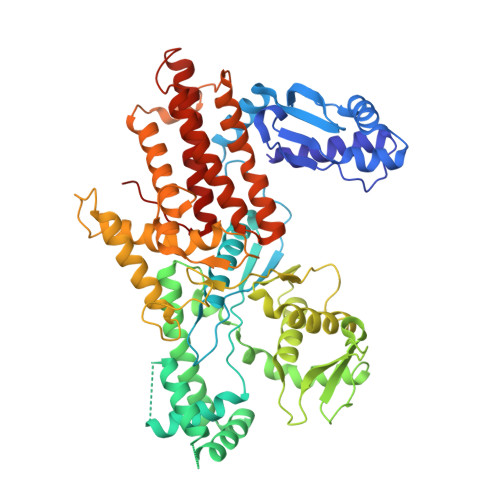
Crystal structure of E. coli arginyl-tRNA synthetase and ligand binding studies revealed key residues in arginine recognition.

Bi, K.Zheng, Y.Gao, F.Dong, J.Wang, J.Wang, Y.Gong, W.

(2014) Protein Cell 5: 151-159

  • DOI: https://doi.org/10.1007/s13238-013-0012-1
  • Primary Citation Related Structures: 
    4OBY

  • PubMed Abstract: 

    The arginyl-tRNA synthetase (ArgRS) catalyzes the esterification reaction between L-arginine and its cognate tRNA(Arg). Previously reported structures of ArgRS shed considerable light on the tRNA recognition mechanism, while the aspect of amino acid binding in ArgRS remains largely unexplored. Here we report the first crystal structure of E. coli ArgRS (eArgRS) complexed with L-arginine, and a series of mutational studies using isothermal titration calorimetry (ITC). Combined with previously reported work on ArgRS, our results elucidated the structural and functional roles of a series of important residues in the active site, which furthered our understanding of this unique enzyme.


  • Organizational Affiliation
    • Laboratory of Non-coding RNA, Institute of Biophysics, Chinese Academy of Sciences, Beijing, 100101, China.

Macromolecule Content 

  • Total Structure Weight: 65.02 kDa 
  • Atom Count: 4,289 
  • Modeled Residue Count: 564 
  • Deposited Residue Count: 578 
  • Unique protein chains: 1

Macromolecules

Find similar proteins by:|  3D Structure
Entity ID: 1
MoleculeChains  Sequence LengthOrganismDetailsImage
Arginine--tRNA ligase578Escherichia coli K-12Mutation(s): 0 
Gene Names: argSb1876JW1865
EC: 6.1.1.19
UniProt
Find proteins for P11875 (Escherichia coli (strain K12))
Explore P11875 
Go to UniProtKB:  P11875
Entity Groups
Sequence Clusters30% Identity50% Identity70% Identity90% Identity95% Identity100% Identity
UniProt GroupP11875
Sequence Annotations
Expand
Reference Sequence

Small Molecules

Ligands 1 Unique
IDChains Name / Formula / InChI Key2D Diagram3D Interactions
ARG

Query on ARG


Download:Ideal Coordinates CCD File
B [auth A]ARGININE
C6 H15 N4 O2
ODKSFYDXXFIFQN-BYPYZUCNSA-O

Experimental Data & Validation

Experimental Data

  • Method: X-RAY DIFFRACTION
  • Resolution: 2.57 Å
  • R-Value Free:  0.278 (Depositor), 0.278 (DCC) 
  • R-Value Work:  0.201 (Depositor), 0.208 (DCC) 
  • R-Value Observed: 0.205 (Depositor) 
Space Group: C 1 2 1
Unit Cell:
Length ( Å )Angle ( ˚ )
a = 118.599α = 90
b = 94.136β = 110.53
c = 62.717γ = 90
Software Package:
Software NamePurpose
PHENIXrefinement
CNSrefinement
HKL-2000data collection
DENZOdata reduction
HKL-2000data scaling
CNSphasing

Structure Validation

View Full Validation Report


Ligand Structure Quality Assessment 


Entry History 

Deposition Data

Revision History  (Full details and data files)

  • Version 1.0: 2014-02-12
    Type: Initial release
  • Version 1.1: 2014-04-02
    Changes: Database references
  • Version 1.2: 2019-07-17
    Changes: Data collection, Refinement description
  • Version 1.3: 2023-09-20
    Changes: Data collection, Database references, Derived calculations, Refinement description