4O6I | pdb_00004o6i

2.0A crystal structure of Lymphocytic Choriomeningitis Virus Nucleoprotein C-terminal Domain


Experimental Data Snapshot

  • Method: X-RAY DIFFRACTION
  • Resolution: 2.00 Å
  • R-Value Free: 
    0.228 (Depositor), 0.234 (DCC) 
  • R-Value Work: 
    0.209 (Depositor), 0.222 (DCC) 
  • R-Value Observed: 
    0.209 (Depositor) 

Starting Model: experimental
View more details

wwPDB Validation 3D Report Full Report

Validation slider image for 4O6I

This is version 1.2 of the entry. See complete history

Literature

Structure of the LCMV nucleoprotein provides a template for understanding arenavirus replication and immunosuppression.

West, B.R.Hastie, K.M.Saphire, E.O.

(2014) Acta Crystallogr D Biol Crystallogr 70: 1764-1769

  • DOI: https://doi.org/10.1107/S1399004714007883
  • Primary Citation Related Structures: 
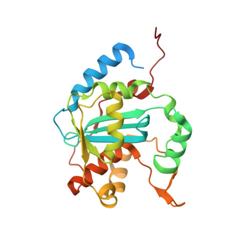
    4O6H, 4O6I

  • PubMed Abstract: 

    The X-ray crystal structure of the Lymphocytic choriomeningitis virus nucleoprotein C-terminal immunosuppressive domain (LCMV NPΔ340) was determined to 2.0 Å resolution. The structure indicates that LCMV NPΔ340, like the other structurally characterized arenaviral nucleoproteins, adopts the fold of an exonuclease. This structure provides a crucial three-dimensional template for functional exploration of the replication and immunosuppression of this prototypic arenavirus.


  • Organizational Affiliation
    • Department of Immunology and Microbial Science, The Scripps Research Institute, La Jolla, CA 92037, USA.

Macromolecule Content 

  • Total Structure Weight: 53.67 kDa 
  • Atom Count: 3,394 
  • Modeled Residue Count: 406 
  • Deposited Residue Count: 466 
  • Unique protein chains: 1

Macromolecules

Find similar proteins by:|  3D Structure
Entity ID: 1
MoleculeChains  Sequence LengthOrganismDetailsImage
Nucleoprotein
A, B
233Lymphocytic choriomeningitis virus (strain Armstrong)Mutation(s): 0 
Gene Names: N
EC: 3.1.13
UniProt
Find proteins for P09992 (Lymphocytic choriomeningitis virus (strain Armstrong))
Explore P09992 
Go to UniProtKB:  P09992
Entity Groups
Sequence Clusters30% Identity50% Identity70% Identity90% Identity95% Identity100% Identity
UniProt GroupP09992
Sequence Annotations
Expand
Reference Sequence

Small Molecules

Ligands 3 Unique
IDChains Name / Formula / InChI Key2D Diagram3D Interactions
IMD

Query on IMD


Download:Ideal Coordinates CCD File
E [auth A]
F [auth A]
G [auth A]
H [auth A]
I [auth A]
E [auth A],
F [auth A],
G [auth A],
H [auth A],
I [auth A],
L [auth B],
M [auth B],
N [auth B],
O [auth B],
P [auth B]
IMIDAZOLE
C3 H5 N2
RAXXELZNTBOGNW-UHFFFAOYSA-O
ZN

Query on ZN


Download:Ideal Coordinates CCD File
C [auth A],
J [auth B]
ZINC ION
Zn
PTFCDOFLOPIGGS-UHFFFAOYSA-N
MG

Query on MG


Download:Ideal Coordinates CCD File
D [auth A],
K [auth B]
MAGNESIUM ION
Mg
JLVVSXFLKOJNIY-UHFFFAOYSA-N

Experimental Data & Validation

Experimental Data

  • Method: X-RAY DIFFRACTION
  • Resolution: 2.00 Å
  • R-Value Free:  0.228 (Depositor), 0.234 (DCC) 
  • R-Value Work:  0.209 (Depositor), 0.222 (DCC) 
  • R-Value Observed: 0.209 (Depositor) 
Space Group: P 65
Unit Cell:
Length ( Å )Angle ( ˚ )
a = 89.962α = 90
b = 89.962β = 90
c = 145.941γ = 120
Software Package:
Software NamePurpose
JBluIce-EPICSdata collection
PHENIXmodel building
PHENIXrefinement
d*TREKdata reduction
d*TREKdata scaling
PHENIXphasing

Structure Validation

View Full Validation Report


Entry History 

Deposition Data

Revision History  (Full details and data files)

  • Version 1.0: 2014-06-11
    Type: Initial release
  • Version 1.1: 2014-07-02
    Changes: Database references
  • Version 1.2: 2023-09-20
    Changes: Data collection, Database references, Derived calculations, Refinement description