4O1F | pdb_00004o1f

Structure of a methyltransferase component in complex with THF involved in O-demethylation


Experimental Data Snapshot

  • Method: X-RAY DIFFRACTION
  • Resolution: 1.80 Å
  • R-Value Free: 
    0.245 (Depositor), 0.237 (DCC) 
  • R-Value Work: 
    0.207 (Depositor) 
  • R-Value Observed: 
    0.209 (Depositor) 

Starting Model: experimental
View more details

wwPDB Validation 3D Report Full Report

Validation slider image for 4O1F

Ligand Structure Quality Assessment 


This is version 1.3 of the entry. See complete history

Literature

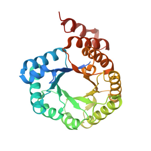
Structures of the methyltransferase component of Desulfitobacterium hafniense DCB-2 O-demethylase shed light on methyltetrahydrofolate formation

Sjuts, H.Dunstan, M.S.Fisher, K.Leys, D.

(2015) Acta Crystallogr D Biol Crystallogr 71: 1900-1908

  • DOI: https://doi.org/10.1107/S1399004715013061
  • Primary Citation Related Structures: 
    4O0Q, 4O1E, 4O1F

  • PubMed Abstract: 

    O-Demethylation by acetogenic or organohalide-respiring bacteria leads to the formation of methyltetrahydrofolate from aromatic methyl ethers. O-Demethylases, which are cobalamin-dependent, three-component enzyme systems, catalyse methyl-group transfers from aromatic methyl ethers to tetrahydrofolate via methylcobalamin intermediates. In this study, crystal structures of the tetrahydrofolate-binding methyltransferase module from a Desulfitobacterium hafniense DCB-2 O-demethylase were determined both in complex with tetrahydrofolate and the product methyltetrahydrofolate. While these structures are similar to previously determined methyltransferase structures, the position of key active-site residues is subtly altered. A strictly conserved Asn is displaced to establish a putative proton-transfer network between the substrate N5 and solvent. It is proposed that this supports the efficient catalysis of methyltetrahydrofolate formation, which is necessary for efficient O-demethylation.


  • Organizational Affiliation
    • Manchester Institute of Biotechnology, Faculty of Life Sciences, University of Manchester, 131 Princess Street, Manchester M1 7DN, England.

Macromolecule Content 

  • Total Structure Weight: 64.4 kDa 
  • Atom Count: 4,399 
  • Modeled Residue Count: 535 
  • Deposited Residue Count: 580 
  • Unique protein chains: 1

Macromolecules

Find similar proteins by:|  3D Structure
Entity ID: 1
MoleculeChains  Sequence LengthOrganismDetailsImage
Dihydropteroate synthase DHPS
A, B
290Desulfitobacterium hafniense DCB-2Mutation(s): 0 
Gene Names: Dhaf_0722
UniProt
Find proteins for B8FW00 (Desulfitobacterium hafniense (strain DSM 10664 / DCB-2))
Explore B8FW00 
Go to UniProtKB:  B8FW00
Entity Groups
Sequence Clusters30% Identity50% Identity70% Identity90% Identity95% Identity100% Identity
UniProt GroupB8FW00
Sequence Annotations
Expand
Reference Sequence

Experimental Data & Validation

Experimental Data

  • Method: X-RAY DIFFRACTION
  • Resolution: 1.80 Å
  • R-Value Free:  0.245 (Depositor), 0.237 (DCC) 
  • R-Value Work:  0.207 (Depositor) 
  • R-Value Observed: 0.209 (Depositor) 
Space Group: P 21 21 2
Unit Cell:
Length ( Å )Angle ( ˚ )
a = 90.19α = 90
b = 118.24β = 90
c = 57.71γ = 90
Software Package:
Software NamePurpose
GDAdata collection
PHASERphasing
REFMACrefinement
xia2data reduction
xia2data scaling

Structure Validation

View Full Validation Report


Ligand Structure Quality Assessment 


Entry History 

Deposition Data

Revision History  (Full details and data files)

  • Version 1.0: 2015-01-21
    Type: Initial release
  • Version 1.1: 2015-09-09
    Changes: Database references
  • Version 1.2: 2015-09-16
    Changes: Database references
  • Version 1.3: 2023-11-08
    Changes: Data collection, Database references, Derived calculations, Refinement description