


An arginine tetrad as mediator of input-dependent and input-independent ATPases in the clock protein KaiC
Pattanayek, R., Xu, Y., Lamichhane, A., Johnson, C.H., Egli, M.(2014) Acta Crystallogr D Biol Crystallogr D70: 1375-1390
Experimental Data Snapshot
(2014) Acta Crystallogr D Biol Crystallogr D70: 1375-1390
Entity ID: 1 | |||||
|---|---|---|---|---|---|
| Molecule | Chains | Sequence Length | Organism | Details | Image |
| Circadian clock protein kinase KaiC | 524 | Thermosynechococcus vestitus BP-1 | Mutation(s): 0 Gene Names: kaiC, tlr0483 EC: 2.7.11.1 (PDB Primary Data), 3.6.4 (UniProt) | 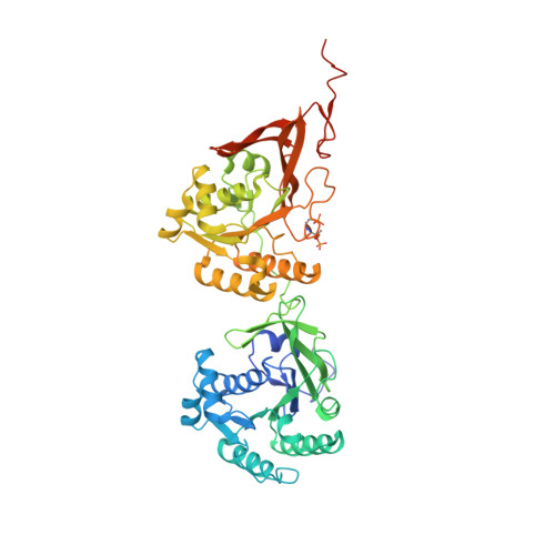 | |
UniProt | |||||
Entity Groups | |||||
| Sequence Clusters | 30% Identity50% Identity70% Identity90% Identity95% Identity100% Identity | ||||
| UniProt Group | Q79V60 | ||||
Sequence AnnotationsExpand | |||||
Reference Sequence | |||||
| Ligands 2 Unique | |||||
|---|---|---|---|---|---|
| ID | Chains | Name / Formula / InChI Key | 2D Diagram | 3D Interactions | |
| ATP Download:Ideal Coordinates CCD File | E [auth A] F [auth A] I [auth B] K [auth B] M [auth C] | ADENOSINE-5'-TRIPHOSPHATE C10 H16 N5 O13 P3 ZKHQWZAMYRWXGA-KQYNXXCUSA-N |  | ||
| MG Download:Ideal Coordinates CCD File | D [auth A] G [auth A] H [auth B] J [auth B] L [auth C] | MAGNESIUM ION Mg JLVVSXFLKOJNIY-UHFFFAOYSA-N |  | ||
| Modified Residues 2 Unique | |||||
|---|---|---|---|---|---|
| ID | Chains | Type | Formula | 2D Diagram | Parent |
| SEP Query on SEP | A, B, C | L-PEPTIDE LINKING | C3 H8 N O6 P |  | SER |
| TPO Query on TPO | A, B, C | L-PEPTIDE LINKING | C4 H10 N O6 P |  | THR |
| Length ( Å ) | Angle ( ˚ ) |
|---|---|
| a = 130.81 | α = 90 |
| b = 195.29 | β = 90 |
| c = 136.64 | γ = 90 |
| Software Name | Purpose |
|---|---|
| HKL-2000 | data collection |
| PHASER | phasing |
| PHENIX | refinement |
| HKL-2000 | data reduction |
| XSCALE | data scaling |


