4NZ3 | pdb_00004nz3

Structure of Vibrio cholerae chitin de-N-acetylase in complex with DI(N-ACETYL-D-GLUCOSAMINE) (CBS) in P 21 21 21


Experimental Data Snapshot

  • Method: X-RAY DIFFRACTION
  • Resolution: 2.11 Å
  • R-Value Free: 
    0.213 (Depositor) 
  • R-Value Work: 
    0.150 (Depositor) 
  • R-Value Observed: 
    0.153 (Depositor) 

Starting Model: experimental
View more details

wwPDB Validation 3D Report Full Report

Validation slider image for 4NZ3

This is version 2.2 of the entry. See complete history

Literature

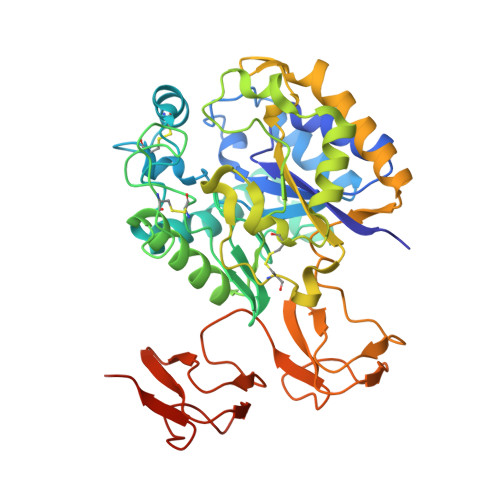
Structural basis of chitin oligosaccharide deacetylation.

Andres, E.Albesa-Jove, D.Biarnes, X.Moerschbacher, B.M.Guerin, M.E.Planas, A.

(2014) Angew Chem Int Ed Engl 53: 6882-6887

  • DOI: https://doi.org/10.1002/anie.201400220
  • Primary Citation Related Structures: 
    4NY2, 4NYU, 4NYY, 4NZ1, 4NZ3, 4NZ4, 4OUI

  • PubMed Abstract: 

    Cell signaling and other biological activities of chitooligosaccharides (COSs) seem to be dependent not only on the degree of polymerization, but markedly on the specific de-N-acetylation pattern. Chitin de-N-acetylases (CDAs) catalyze the hydrolysis of the acetamido group in GlcNAc residues of chitin, chitosan, and COS. A major challenge is to understand how CDAs specifically define the distribution of GlcNAc and GlcNH2 moieties in the oligomeric chain. We report the crystal structure of the Vibrio cholerae CDA in four relevant states of its catalytic cycle. The two enzyme complexes with chitobiose and chitotriose represent the first 3D structures of a CDA with its natural substrates in a productive mode for catalysis, thereby unraveling an induced-fit mechanism with a significant conformational change of a loop closing the active site. We propose that the deacetylation pattern exhibited by different CDAs is governed by critical loops that shape and differentially block accessible subsites in the binding cleft of CE4 enzymes.


  • Organizational Affiliation
    • Laboratory of Biochemistry, Institut Químic de Sarrià, Universitat Ramon Llull, Via Augusta 390, 08017 Barcelona (Spain).

Macromolecule Content 

  • Total Structure Weight: 95.19 kDa 
  • Atom Count: 7,295 
  • Modeled Residue Count: 808 
  • Deposited Residue Count: 838 
  • Unique protein chains: 1

Macromolecules

Find similar proteins by:|  3D Structure
Entity ID: 1
MoleculeChains  Sequence LengthOrganismDetailsImage
Deacetylase DA1
A, B
419Vibrio cholerae O1 str. NHCC-010FMutation(s): 1 
EC: 3.5.1.41

Oligosaccharides

Help  
Entity ID: 2
MoleculeChains Length2D Diagram GlycosylationD Interactions
2-acetamido-2-deoxy-beta-D-glucopyranose-(1-4)-2-acetamido-2-deoxy-beta-D-glucopyranose
C, D
2N/A
Glycosylation Resources
GlyTouCan: G42666HT
GlyCosmos: G42666HT
GlyGen: G42666HT

Small Molecules

Ligands 3 Unique
IDChains Name / Formula / InChI Key2D Diagram3D Interactions
ZN

Query on ZN


Download:Ideal Coordinates CCD File
E [auth A],
K [auth B]
ZINC ION
Zn
PTFCDOFLOPIGGS-UHFFFAOYSA-N
EDO

Query on EDO


Download:Ideal Coordinates CCD File
G [auth A]
H [auth A]
I [auth A]
J [auth A]
M [auth B]
G [auth A],
H [auth A],
I [auth A],
J [auth A],
M [auth B],
N [auth B]
1,2-ETHANEDIOL
C2 H6 O2
LYCAIKOWRPUZTN-UHFFFAOYSA-N
MG

Query on MG


Download:Ideal Coordinates CCD File
F [auth A],
L [auth B]
MAGNESIUM ION
Mg
JLVVSXFLKOJNIY-UHFFFAOYSA-N

Experimental Data & Validation

Experimental Data

  • Method: X-RAY DIFFRACTION
  • Resolution: 2.11 Å
  • R-Value Free:  0.213 (Depositor) 
  • R-Value Work:  0.150 (Depositor) 
  • R-Value Observed: 0.153 (Depositor) 
Space Group: P 21 21 21
Unit Cell:
Length ( Å )Angle ( ˚ )
a = 63.684α = 90
b = 98.142β = 90
c = 152.988γ = 90
Software Package:
Software NamePurpose
GDAdata collection
PHASERphasing
PHENIXrefinement
XDSdata reduction
XDSdata scaling

Structure Validation

View Full Validation Report


Entry History 

Deposition Data

Revision History  (Full details and data files)

  • Version 1.0: 2014-08-13
    Type: Initial release
  • Version 1.1: 2014-11-26
    Changes: Structure summary
  • Version 2.0: 2020-07-29
    Type: Remediation
    Reason: Carbohydrate remediation
    Changes: Atomic model, Data collection, Database references, Derived calculations, Structure summary
  • Version 2.1: 2023-11-08
    Changes: Data collection, Database references, Refinement description, Structure summary
  • Version 2.2: 2024-11-13
    Changes: Structure summary