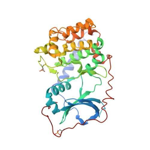
4NTS

Apo structure of the catalytic subunit of cAMP-dependent protein kinase


Experimental Data Snapshot

  • Method: X-RAY DIFFRACTION
  • Resolution: 2.90 Å
  • R-Value Free: 0.292 
  • R-Value Work: 0.244 
  • R-Value Observed: 0.247 

Starting Model: experimental
View more details

wwPDB Validation   3D Report Full Report


Ligand Structure Quality Assessment 


This is version 1.3 of the entry. See complete history


Literature

Molecular Features of Product Release for the PKA Catalytic Cycle.

Bastidas, A.C.Wu, J.Taylor, S.S.

(2015) Biochemistry 54: 2-10

  • DOI: https://doi.org/10.1021/bi500684c
  • Primary Citation of Related Structures:  
    4NTS, 4NTT

  • PubMed Abstract: 

    Although ADP release is the rate limiting step in product turnover by protein kinase A, the steps and motions involved in this process are not well resolved. Here we report the apo and ADP bound structures of the myristylated catalytic subunit of PKA at 2.9 and 3.5 Å resolution, respectively. The ADP bound structure adopts a conformation that does not conform to the previously characterized open, closed, or intermediate states. In the ADP bound structure, the C-terminal tail and Gly-rich loop are more closed than in the open state adopted in the apo structure but are also much more open than the intermediate or closed conformations. Furthermore, ADP binds at the active site with only one magnesium ion, termed Mg2 from previous structures. These structures thus support a model where ADP release proceeds through release of the substrate and Mg1 followed by lifting of the Gly-rich loop and disengagement of the C-terminal tail. Coupling of these two structural elements with the release of the first metal ion fills in a key step in the catalytic cycle that has been missing and supports an ensemble of correlated conformational states that mediate the full catalytic cycle for a protein kinase.


  • Organizational Affiliation

    Department of Pharmacology, University of California, San Diego , San Diego, California 92093, United States.


Macromolecules
Find similar proteins by:  (by identity cutoff)  |  3D Structure
Entity ID: 1
MoleculeChains Sequence LengthOrganismDetailsImage
cAMP-dependent protein kinase catalytic subunit alpha
A, B
350Mus musculusMutation(s): 1 
Gene Names: PrkacaPkaca
EC: 2.7.11.11
UniProt
Find proteins for P05132 (Mus musculus)
Explore P05132 
Go to UniProtKB:  P05132
Entity Groups  
Sequence Clusters30% Identity50% Identity70% Identity90% Identity95% Identity100% Identity
UniProt GroupP05132
Sequence Annotations
Expand
  • Reference Sequence
Small Molecules
Ligands 1 Unique
IDChains Name / Formula / InChI Key2D Diagram3D Interactions
MYR
Query on MYR

Download Ideal Coordinates CCD File 
C [auth A]MYRISTIC ACID
C14 H28 O2
TUNFSRHWOTWDNC-UHFFFAOYSA-N
Modified Residues  2 Unique
IDChains TypeFormula2D DiagramParent
SEP
Query on SEP
A, B
L-PEPTIDE LINKINGC3 H8 N O6 PSER
TPO
Query on TPO
A, B
L-PEPTIDE LINKINGC4 H10 N O6 PTHR
Experimental Data & Validation

Experimental Data

  • Method: X-RAY DIFFRACTION
  • Resolution: 2.90 Å
  • R-Value Free: 0.292 
  • R-Value Work: 0.244 
  • R-Value Observed: 0.247 
  • Space Group: P 1 21 1
Unit Cell:
Length ( Å )Angle ( ˚ )
a = 50.96α = 90
b = 142.61β = 103.65
c = 63.16γ = 90
Software Package:
Software NamePurpose
REFMACrefinement

Structure Validation

View Full Validation Report


Ligand Structure Quality Assessment 


Entry History 

Deposition Data

Revision History  (Full details and data files)

  • Version 1.0: 2014-10-15
    Type: Initial release
  • Version 1.1: 2015-01-28
    Changes: Database references
  • Version 1.2: 2023-09-20
    Changes: Data collection, Database references, Derived calculations, Refinement description
  • Version 1.3: 2024-11-20
    Changes: Structure summary