4NO7

Human Glucokinase in complex with a nanomolar activator.


Experimental Data Snapshot

  • Method: X-RAY DIFFRACTION
  • Resolution: 1.70 Å
  • R-Value Free: 0.223 
  • R-Value Work: 0.190 
  • R-Value Observed: 0.191 

wwPDB Validation   3D Report Full Report


Ligand Structure Quality Assessment 


This is version 1.4 of the entry. See complete history


Literature

The active conformation of human glucokinase is not altered by allosteric activators.

Petit, P.Antoine, M.Ferry, G.Boutin, J.A.Lagarde, A.Gluais, L.Vincentelli, R.Vuillard, L.

(2011) Acta Crystallogr D Biol Crystallogr 67: 929-935

  • DOI: https://doi.org/10.1107/S0907444911036729
  • Primary Citation of Related Structures:  
    3F9M, 3FGU, 3ID8, 3IDH, 4NO7

  • PubMed Abstract: 

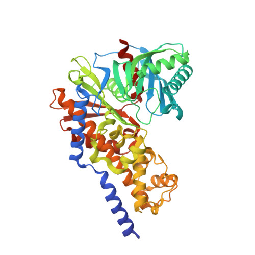
    Glucokinase (GK) catalyses the formation of glucose 6-phosphate from glucose and ATP. A specific feature of GK amongst hexokinases is that it can cycle between active and inactive conformations as a function of glucose concentration, resulting in a unique positive kinetic cooperativity with glucose, which turns GK into a unique key sensor of glucose metabolism, notably in the pancreas. GK is a target of antidiabetic drugs aimed at the activation of GK activity, leading to insulin secretion. Here, the first structures of a GK-glucose complex without activator, of GK-glucose-AMP-PNP and of GK-glucose-AMP-PNP with a bound activator are reported. All these structures are extremely similar, thus demonstrating that binding of GK activators does not result in conformational changes of the active protein but in stabilization of the active form of GK.


  • Organizational Affiliation

    BioXtal, PX Unit, c/o AFMB, UMR 6098, Case 932, 163 Avenue de Luminy, 13288 Marseille CEDEX 09, France.


Macromolecules
Find similar proteins by:  (by identity cutoff)  |  3D Structure
Entity ID: 1
MoleculeChains Sequence LengthOrganismDetailsImage
Glucokinase470Homo sapiensMutation(s): 0 
Gene Names: GCK
EC: 2.7.1.2
UniProt & NIH Common Fund Data Resources
Find proteins for P35557 (Homo sapiens)
Explore P35557 
Go to UniProtKB:  P35557
PHAROS:  P35557
GTEx:  ENSG00000106633 
Entity Groups  
Sequence Clusters30% Identity50% Identity70% Identity90% Identity95% Identity100% Identity
UniProt GroupP35557
Sequence Annotations
Expand
  • Reference Sequence
Small Molecules
Ligands 2 Unique
IDChains Name / Formula / InChI Key2D Diagram3D Interactions
2N8
Query on 2N8

Download Ideal Coordinates CCD File 
C [auth A](2R)-2-[3-chloro-4-(methylsulfonyl)phenyl]-3-[(1R)-3-oxocyclopentyl]-N-(pyrazin-2-yl)propanamide
C19 H20 Cl N3 O4 S
XEANIURBPHCHMG-SWLSCSKDSA-N
GLC
Query on GLC

Download Ideal Coordinates CCD File 
B [auth A]alpha-D-glucopyranose
C6 H12 O6
WQZGKKKJIJFFOK-DVKNGEFBSA-N
Binding Affinity Annotations 
IDSourceBinding Affinity
2N8 BindingDB:  4NO7 Kd: 830 (nM) from 1 assay(s)
EC50: min: 1420, max: 6320 (nM) from 2 assay(s)
Experimental Data & Validation

Experimental Data

  • Method: X-RAY DIFFRACTION
  • Resolution: 1.70 Å
  • R-Value Free: 0.223 
  • R-Value Work: 0.190 
  • R-Value Observed: 0.191 
  • Space Group: P 21 21 21
Unit Cell:
Length ( Å )Angle ( ˚ )
a = 66.3α = 90
b = 81.3β = 90
c = 85.5γ = 90
Software Package:
Software NamePurpose
REFMACrefinement
CNSrefinement
HKL-2000data collection
MOSFLMdata reduction
SCALAdata scaling
CNSphasing

Structure Validation

View Full Validation Report


Ligand Structure Quality Assessment 


Entry History 

Deposition Data

Revision History  (Full details and data files)

  • Version 1.0: 2014-10-29
    Type: Initial release
  • Version 1.1: 2018-04-18
    Changes: Data collection, Database references
  • Version 1.2: 2019-07-17
    Changes: Data collection, Refinement description
  • Version 1.3: 2020-07-29
    Type: Remediation
    Reason: Carbohydrate remediation
    Changes: Data collection, Database references, Derived calculations, Structure summary
  • Version 1.4: 2023-09-20
    Changes: Data collection, Database references, Refinement description, Structure summary