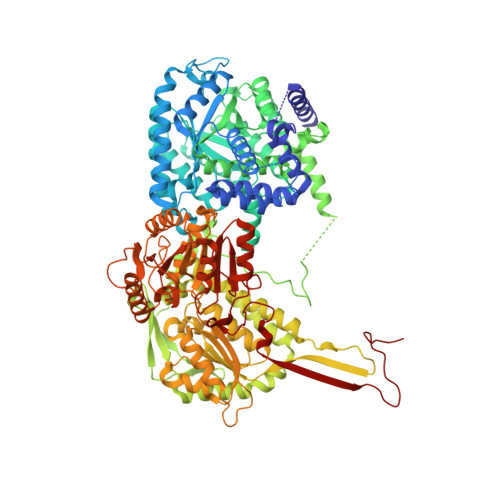
4NMF

Crystal structure of proline utilization A (PutA) from Geobacter sulfurreducens PCA inactivated by N-propargylglycine and complexed with menadione bisulfite


Experimental Data Snapshot

  • Method: X-RAY DIFFRACTION
  • Resolution: 1.95 Å
  • R-Value Free: 0.203 
  • R-Value Work: 0.173 
  • R-Value Observed: 0.174 

Starting Model: experimental
View more details

wwPDB Validation   3D Report Full Report


Ligand Structure Quality Assessment 


This is version 1.2 of the entry. See complete history


Literature

Structures of the PutA peripheral membrane flavoenzyme reveal a dynamic substrate-channeling tunnel and the quinone-binding site.

Singh, H.Arentson, B.W.Becker, D.F.Tanner, J.J.

(2014) Proc Natl Acad Sci U S A 111: 3389-3394

  • DOI: https://doi.org/10.1073/pnas.1321621111
  • Primary Citation of Related Structures:  
    4NM9, 4NMA, 4NMB, 4NMC, 4NMD, 4NME, 4NMF

  • PubMed Abstract: 

    Proline utilization A (PutA) proteins are bifunctional peripheral membrane flavoenzymes that catalyze the oxidation of L-proline to L-glutamate by the sequential activities of proline dehydrogenase and aldehyde dehydrogenase domains. Located at the inner membrane of Gram-negative bacteria, PutAs play a major role in energy metabolism by coupling the oxidation of proline imported from the environment to the reduction of membrane-associated quinones. Here, we report seven crystal structures of the 1,004-residue PutA from Geobacter sulfurreducens, along with determination of the protein oligomeric state by small-angle X-ray scattering and kinetic characterization of substrate channeling and quinone reduction. The structures reveal an elaborate and dynamic tunnel system featuring a 75-Å-long tunnel that links the two active sites and six smaller tunnels that connect the main tunnel to the bulk medium. The locations of these tunnels and their responses to ligand binding and flavin reduction suggest hypotheses about how proline, water, and quinones enter the tunnel system and where L-glutamate exits. Kinetic measurements show that glutamate production from proline occurs without a lag phase, consistent with substrate channeling and implying that the observed tunnel is functionally relevant. Furthermore, the structure of reduced PutA complexed with menadione bisulfite reveals the elusive quinone-binding site. The benzoquinone binds within 4.0 Å of the flavin si face, consistent with direct electron transfer. The location of the quinone site implies that the concave surface of the PutA dimer approaches the membrane. Altogether, these results provide insight into how PutAs couple proline oxidation to quinone reduction.


  • Organizational Affiliation

    Departments of Chemistry and Biochemistry, University of Missouri, Columbia, MO 65211.


Macromolecules
Find similar proteins by:  (by identity cutoff)  |  3D Structure
Entity ID: 1
MoleculeChains Sequence LengthOrganismDetailsImage
Proline dehydrogenase and Delta-1-pyrroline-5-carboxylate dehydrogenase
A, B
1,005Geobacter sulfurreducens PCAMutation(s): 0 
Gene Names: putAGSU3395
EC: 1.5.99.8 (PDB Primary Data), 1.5.1.12 (PDB Primary Data), 1.2.1.88 (PDB Primary Data)
UniProt
Find proteins for Q746X3 (Geobacter sulfurreducens (strain ATCC 51573 / DSM 12127 / PCA))
Explore Q746X3 
Go to UniProtKB:  Q746X3
Entity Groups  
Sequence Clusters30% Identity50% Identity70% Identity90% Identity95% Identity100% Identity
UniProt GroupQ746X3
Sequence Annotations
Expand
  • Reference Sequence
Small Molecules
Ligands 4 Unique
IDChains Name / Formula / InChI Key2D Diagram3D Interactions
P5F
Query on P5F

Download Ideal Coordinates CCD File 
C [auth A],
J [auth B]
N-propargylglycine-modified flavin adenine dinucleotide
C32 H40 N10 O17 P2
IRSTVKNHNHEQIM-RDJFLKOLSA-N
2LB
Query on 2LB

Download Ideal Coordinates CCD File 
L [auth B](2S)-2-methyl-1,4-dioxo-1,2,3,4-tetrahydronaphthalene-2-sulfonic acid
C11 H10 O5 S
WIXFIQKTHUVFDI-NSHDSACASA-N
2L3
Query on 2L3

Download Ideal Coordinates CCD File 
D [auth A],
K [auth B]
(2R)-2-methyl-1,4-dioxo-1,2,3,4-tetrahydronaphthalene-2-sulfonic acid
C11 H10 O5 S
WIXFIQKTHUVFDI-LLVKDONJSA-N
EDO
Query on EDO

Download Ideal Coordinates CCD File 
E [auth A]
F [auth A]
G [auth A]
H [auth A]
I [auth A]
E [auth A],
F [auth A],
G [auth A],
H [auth A],
I [auth A],
M [auth B],
N [auth B],
O [auth B]
1,2-ETHANEDIOL
C2 H6 O2
LYCAIKOWRPUZTN-UHFFFAOYSA-N
Experimental Data & Validation

Experimental Data

  • Method: X-RAY DIFFRACTION
  • Resolution: 1.95 Å
  • R-Value Free: 0.203 
  • R-Value Work: 0.173 
  • R-Value Observed: 0.174 
  • Space Group: P 21 21 21
Unit Cell:
Length ( Å )Angle ( ˚ )
a = 95.594α = 90
b = 151.741β = 90
c = 176.606γ = 90
Software Package:
Software NamePurpose
SCALAdata scaling
PHENIXrefinement
PDB_EXTRACTdata extraction
XDSdata reduction
PHENIXphasing

Structure Validation

View Full Validation Report


Ligand Structure Quality Assessment 


Entry History 

Deposition Data

Revision History  (Full details and data files)

  • Version 1.0: 2014-02-19
    Type: Initial release
  • Version 1.1: 2014-03-19
    Changes: Database references
  • Version 1.2: 2023-09-20
    Changes: Advisory, Data collection, Database references, Derived calculations, Refinement description