4NI0

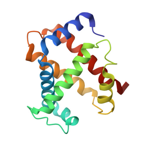
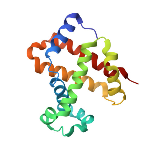
Quaternary R3 CO-liganded hemoglobin structure in complex with a thiol containing compound


Experimental Data Snapshot

  • Method: X-RAY DIFFRACTION
  • Resolution: 2.15 Å
  • R-Value Free: 0.259 
  • R-Value Work: 0.228 
  • R-Value Observed: 0.228 

Starting Model: experimental
View more details

wwPDB Validation   3D Report Full Report


Ligand Structure Quality Assessment 


This is version 1.2 of the entry. See complete history


Literature

Identification of a Small Molecule that Increases Hemoglobin Oxygen Affinity and Reduces SS Erythrocyte Sickling.

Nakagawa, A.Lui, F.E.Wassaf, D.Yefidoff-Freedman, R.Casalena, D.Palmer, M.A.Meadows, J.Mozzarelli, A.Ronda, L.Abdulmalik, O.Bloch, K.D.Safo, M.K.Zapol, W.M.

(2014) ACS Chem Biol 9: 2318-2325

  • DOI: https://doi.org/10.1021/cb500230b
  • Primary Citation of Related Structures:  
    4NI0, 4NI1

  • PubMed Abstract: 

    Small molecules that increase the oxygen affinity of human hemoglobin may reduce sickling of red blood cells in patients with sickle cell disease. We screened 38,700 compounds using small molecule microarrays and identified 427 molecules that bind to hemoglobin. We developed a high-throughput assay for evaluating the ability of the 427 small molecules to modulate the oxygen affinity of hemoglobin. We identified a novel allosteric effector of hemoglobin, di(5-(2,3-dihydro-1,4-benzodioxin-2-yl)-4H-1,2,4-triazol-3-yl)disulfide (TD-1). TD-1 induced a greater increase in oxygen affinity of human hemoglobin in solution and in red blood cells than did 5-hydroxymethyl-2-furfural (5-HMF), N-ethylmaleimide (NEM), or diformamidine disulfide. The three-dimensional structure of hemoglobin complexed with TD-1 revealed that monomeric units of TD-1 bound covalently to β-Cys93 and β-Cys112, as well as noncovalently to the central water cavity of the hemoglobin tetramer. The binding of TD-1 to hemoglobin stabilized the relaxed state (R3-state) of hemoglobin. TD-1 increased the oxygen affinity of sickle hemoglobin and inhibited in vitro hypoxia-induced sickling of red blood cells in patients with sickle cell disease without causing hemolysis. Our study indicates that TD-1 represents a novel lead molecule for the treatment of patients with sickle cell disease.


  • Organizational Affiliation

    Anesthesia Center for Critical Care Research, Department of Anesthesia, Critical Care, and Pain Medicine, Massachusetts General Hospital and Harvard Medical School, 55 Fruit Street, Boston, Massachusetts 02114, United States.


Macromolecules
Find similar proteins by:  (by identity cutoff)  |  3D Structure
Entity ID: 1
MoleculeChains Sequence LengthOrganismDetailsImage
Hemoglobin subunit alpha141Homo sapiensMutation(s): 0 
UniProt & NIH Common Fund Data Resources
Find proteins for P69905 (Homo sapiens)
Explore P69905 
Go to UniProtKB:  P69905
PHAROS:  P69905
Entity Groups  
Sequence Clusters30% Identity50% Identity70% Identity90% Identity95% Identity100% Identity
UniProt GroupP69905
Sequence Annotations
Expand
  • Reference Sequence
Find similar proteins by:  (by identity cutoff)  |  3D Structure
Entity ID: 2
MoleculeChains Sequence LengthOrganismDetailsImage
Hemoglobin subunit beta146Homo sapiensMutation(s): 0 
UniProt & NIH Common Fund Data Resources
Find proteins for P68871 (Homo sapiens)
Explore P68871 
Go to UniProtKB:  P68871
PHAROS:  P68871
GTEx:  ENSG00000244734 
Entity Groups  
Sequence Clusters30% Identity50% Identity70% Identity90% Identity95% Identity100% Identity
UniProt GroupP68871
Sequence Annotations
Expand
  • Reference Sequence
Small Molecules
Ligands 5 Unique
IDChains Name / Formula / InChI Key2D Diagram3D Interactions
HEM
Query on HEM

Download Ideal Coordinates CCD File 
D [auth A],
L [auth B]
PROTOPORPHYRIN IX CONTAINING FE
C34 H32 Fe N4 O4
KABFMIBPWCXCRK-RGGAHWMASA-L
2P3
Query on 2P3

Download Ideal Coordinates CCD File 
E [auth A],
F [auth A],
M [auth B],
N [auth B]
5-[(2S)-2,3-dihydro-1,4-benzodioxin-2-yl]-2,4-dihydro-3H-1,2,4-triazole-3-thione
C10 H9 N3 O2 S
WDUGNJIRKJHDNF-MRVPVSSYSA-N
PO4
Query on PO4

Download Ideal Coordinates CCD File 
J [auth A],
O [auth B],
P [auth B]
PHOSPHATE ION
O4 P
NBIIXXVUZAFLBC-UHFFFAOYSA-K
MBN
Query on MBN

Download Ideal Coordinates CCD File 
G [auth A],
H [auth A],
I [auth A]
TOLUENE
C7 H8
YXFVVABEGXRONW-UHFFFAOYSA-N
CMO
Query on CMO

Download Ideal Coordinates CCD File 
C [auth A],
K [auth B]
CARBON MONOXIDE
C O
UGFAIRIUMAVXCW-UHFFFAOYSA-N
Experimental Data & Validation

Experimental Data

  • Method: X-RAY DIFFRACTION
  • Resolution: 2.15 Å
  • R-Value Free: 0.259 
  • R-Value Work: 0.228 
  • R-Value Observed: 0.228 
  • Space Group: P 41 2 2
Unit Cell:
Length ( Å )Angle ( ˚ )
a = 62.0636α = 90
b = 62.0636β = 90
c = 173.8816γ = 90
Software Package:
Software NamePurpose
CrystalCleardata collection
CNSrefinement
d*TREKdata reduction
d*TREKdata scaling
CNSphasing

Structure Validation

View Full Validation Report


Ligand Structure Quality Assessment 


Entry History 

Deposition Data

Revision History  (Full details and data files)

  • Version 1.0: 2014-08-20
    Type: Initial release
  • Version 1.1: 2014-10-29
    Changes: Database references
  • Version 1.2: 2023-09-20
    Changes: Data collection, Database references, Derived calculations, Refinement description