4NHA

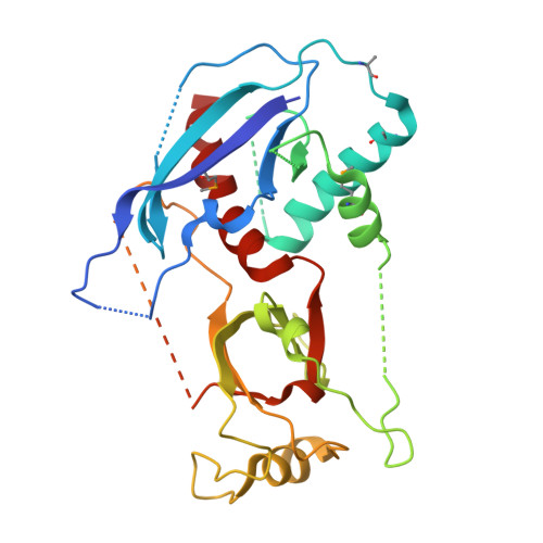
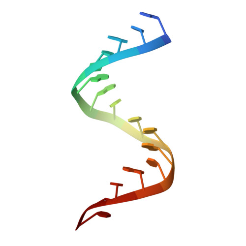
Structure of human Dicer Platform-PAZ-Connector Helix cassette in complex with 16-mer siRNA having 5'-p and UU-3' ends (3.4 Angstrom resolution)


Experimental Data Snapshot

  • Method: X-RAY DIFFRACTION
  • Resolution: 3.40 Å
  • R-Value Free: 0.317 
  • R-Value Work: 0.277 
  • R-Value Observed: 0.281 

wwPDB Validation   3D Report Full Report


This is version 1.4 of the entry. See complete history


Literature

A Phosphate-Binding Pocket within the Platform-PAZ-Connector Helix Cassette of Human Dicer.

Tian, Y.Simanshu, D.K.Ma, J.B.Park, J.E.Heo, I.Kim, V.N.Patel, D.J.

(2014) Mol Cell 53: 606-616

  • DOI: https://doi.org/10.1016/j.molcel.2014.01.003
  • Primary Citation of Related Structures:  
    4NGB, 4NGC, 4NGD, 4NGF, 4NGG, 4NH3, 4NH5, 4NH6, 4NHA

  • PubMed Abstract: 

    We have solved two families of crystal structures of the human Dicer "platform-PAZ-connector helix" cassette in complex with small interfering RNAs (siRNAs). The structures possess two adjacently positioned pockets: a 2 nt 3'-overhang-binding pocket within the PAZ domain (3' pocket) and a phosphate-binding pocket within the platform domain (phosphate pocket). One family of complexes contains a knob-like α-helical protrusion, designated "hDicer-specific helix," that separates the two pockets and orients the bound siRNA away from the surface of Dicer, which could be indicative of a product release/transfer state. In the second complex, the helical protrusion is melted/disordered and the bound siRNA is aligned toward the surface of Dicer, suggestive of a cleavage-competent state. These structures allow us to propose that the transition from the cleavage-competent to the postulated product release/transfer state may involve release of the 5'-phosphate from the phosphate pocket while retaining the 3' overhang in the 3' pocket.


  • Organizational Affiliation

    Structural Biology Program, Memorial Sloan-Kettering Cancer Center, New York, NY 10065, USA.


Macromolecules

Find similar proteins by:  (by identity cutoff)  |  3D Structure
Entity ID: 1
MoleculeChains Sequence LengthOrganismDetailsImage
Endoribonuclease Dicer302Homo sapiensMutation(s): 0 
Gene Names: DICERDICER1HERNAKIAA0928
EC: 3.1.26 (PDB Primary Data), 3.1.26.3 (UniProt)
UniProt & NIH Common Fund Data Resources
Find proteins for Q9UPY3 (Homo sapiens)
Explore Q9UPY3 
Go to UniProtKB:  Q9UPY3
PHAROS:  Q9UPY3
GTEx:  ENSG00000100697 
Entity Groups  
Sequence Clusters30% Identity50% Identity70% Identity90% Identity95% Identity100% Identity
UniProt GroupQ9UPY3
Sequence Annotations
Expand
  • Reference Sequence

Find similar nucleic acids by:  Sequence   |   3D Structure  

Entity ID: 2
MoleculeChains LengthOrganismImage
5'-R(P*GP*CP*GP*UP*UP*GP*GP*CP*CP*AP*AP*CP*GP*CP*UP*U)-3'16N/A
Sequence Annotations
Expand
  • Reference Sequence
Small Molecules
Modified Residues  1 Unique
IDChains TypeFormula2D DiagramParent
MSE
Query on MSE
A
L-PEPTIDE LINKINGC5 H11 N O2 SeMET
Experimental Data & Validation

Experimental Data

  • Method: X-RAY DIFFRACTION
  • Resolution: 3.40 Å
  • R-Value Free: 0.317 
  • R-Value Work: 0.277 
  • R-Value Observed: 0.281 
  • Space Group: I 41 2 2
Unit Cell:
Length ( Å )Angle ( ˚ )
a = 174.469α = 90
b = 174.469β = 90
c = 74.757γ = 90
Software Package:
Software NamePurpose
DENZOdata reduction
SCALEPACKdata scaling
PHENIXrefinement
PDB_EXTRACTdata extraction
HKL-2000data reduction
SHARPphasing

Structure Validation

View Full Validation Report


Entry History 

Deposition Data

Revision History  (Full details and data files)

  • Version 1.0: 2014-03-05
    Type: Initial release
  • Version 1.1: 2014-03-12
    Changes: Database references
  • Version 1.2: 2017-11-15
    Changes: Refinement description
  • Version 1.3: 2018-01-24
    Changes: Structure summary
  • Version 1.4: 2024-11-20
    Changes: Data collection, Database references, Derived calculations, Structure summary