4NG2 | pdb_00004ng2

Crystal structure of LasR LBD-QslA complex from Pseudomonas aeruginosa


Experimental Data Snapshot

  • Method: X-RAY DIFFRACTION
  • Resolution: 2.41 Å
  • R-Value Free: 
    0.277 (Depositor), 0.276 (DCC) 
  • R-Value Work: 
    0.231 (Depositor), 0.231 (DCC) 
  • R-Value Observed: 
    0.234 (Depositor) 

Starting Model: experimental
View more details

wwPDB Validation 3D Report Full Report

Validation slider image for 4NG2

Ligand Structure Quality Assessment 


This is version 1.2 of the entry. See complete history

Literature

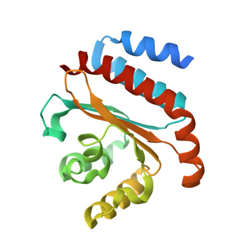
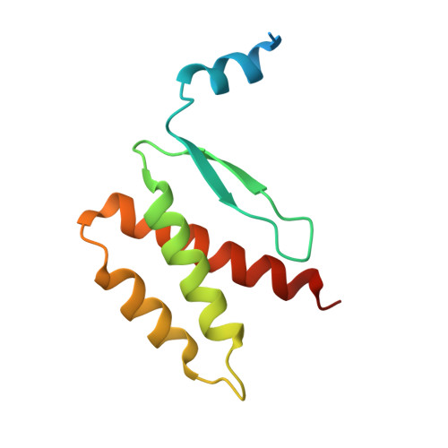
QsIA disrupts LasR dimerization in antiactivation of bacterial quorum sensing

Fan, H.Dong, Y.Wu, D.H.Bowler, M.W.Zhang, L.Song, H.

(2013) Proc Natl Acad Sci U S A 110: 20765-20770

  • DOI: https://doi.org/10.1073/pnas.1314415110
  • Primary Citation Related Structures: 
    4NG2

  • PubMed Abstract: 

    The human pathogen Pseudomonas aeruginosa coordinates the expression of virulence factors by using quorum sensing (QS), a signaling cascade triggered by the QS signal molecule and its receptor, a member of the LuxR family of QS transcriptional factors (LasR). The QS threshold and response in P. aeruginosa is defined by a QS LasR-specific antiactivator (QslA), which binds to LasR and prevents it from binding to its target promoter. However, how QslA binds to LasR and regulates its DNA binding activity in QS remains elusive. Here we report the crystal structure of QslA in complex with the N-terminal ligand binding domain of LasR. QsIA exists as a functional dimer to interact with the LasR ligand binding domain. Further analysis shows that QsIA binding occupies the LasR dimerization interface and consequently disrupts LasR dimerization, thereby preventing LasR from binding to its target DNA and disturbing normal QS. Our findings provide a structural model for understanding the QslA-mediated antiactivation mechanism in QS through protein-protein interaction.


  • Organizational Affiliation
    • Institute of Molecular and Cell Biology, Singapore 138673.

Macromolecule Content 

  • Total Structure Weight: 185.89 kDa 
  • Atom Count: 11,158 
  • Modeled Residue Count: 1,368 
  • Deposited Residue Count: 1,640 
  • Unique protein chains: 2

Macromolecules

Find similar proteins by:|  3D Structure
Entity ID: 1
MoleculeChains  Sequence LengthOrganismDetailsImage
Transcriptional activator protein LasR
A, B, C, D
184Pseudomonas aeruginosa PAO1Mutation(s): 0 
Gene Names: lasR
UniProt
Find proteins for P25084 (Pseudomonas aeruginosa (strain ATCC 15692 / DSM 22644 / CIP 104116 / JCM 14847 / LMG 12228 / 1C / PRS 101 / PAO1))
Explore P25084 
Go to UniProtKB:  P25084
Entity Groups
Sequence Clusters30% Identity50% Identity70% Identity90% Identity95% Identity100% Identity
UniProt GroupP25084
Sequence Annotations
Expand
Reference Sequence
Find similar proteins by:|  3D Structure
Entity ID: 2
MoleculeChains  Sequence LengthOrganismDetailsImage
Uncharacterized protein
E, F, G, H, I
E, F, G, H, I, J, K, L
113Pseudomonas aeruginosa PAO1Mutation(s): 0 
Gene Names: PA1244
UniProt
Find proteins for Q9I494 (Pseudomonas aeruginosa (strain ATCC 15692 / DSM 22644 / CIP 104116 / JCM 14847 / LMG 12228 / 1C / PRS 101 / PAO1))
Explore Q9I494 
Go to UniProtKB:  Q9I494
Entity Groups
Sequence Clusters30% Identity50% Identity70% Identity90% Identity95% Identity100% Identity
UniProt GroupQ9I494
Sequence Annotations
Expand
Reference Sequence

Experimental Data & Validation

Experimental Data

  • Method: X-RAY DIFFRACTION
  • Resolution: 2.41 Å
  • R-Value Free:  0.277 (Depositor), 0.276 (DCC) 
  • R-Value Work:  0.231 (Depositor), 0.231 (DCC) 
  • R-Value Observed: 0.234 (Depositor) 
Space Group: P 21 21 2
Unit Cell:
Length ( Å )Angle ( ˚ )
a = 163.507α = 90
b = 185.887β = 90
c = 56.106γ = 90
Software Package:
Software NamePurpose
ADSCdata collection
PHENIXmodel building
PHENIXrefinement
MOSFLMdata reduction
SCALAdata scaling
PHENIXphasing

Structure Validation

View Full Validation Report


Ligand Structure Quality Assessment 


Entry History 

Deposition Data

Revision History  (Full details and data files)

  • Version 1.0: 2013-12-18
    Type: Initial release
  • Version 1.1: 2015-09-23
    Changes: Database references
  • Version 1.2: 2023-11-08
    Changes: Data collection, Database references, Derived calculations, Refinement description