4NCN | pdb_00004ncn

Crystal structure of eukaryotic translation initiation factor eIF5B (517-858) from Chaetomium thermophilum in complex with GTP


Experimental Data Snapshot

  • Method: X-RAY DIFFRACTION
  • Resolution: 1.87 Å
  • R-Value Free: 
    0.204 (Depositor), 0.205 (DCC) 
  • R-Value Work: 
    0.164 (Depositor), 0.166 (DCC) 
  • R-Value Observed: 
    0.166 (Depositor) 

Starting Model: experimental
View more details

wwPDB Validation 3D Report Full Report

Validation slider image for 4NCN

Ligand Structure Quality Assessment 


This is version 1.3 of the entry. See complete history

Literature

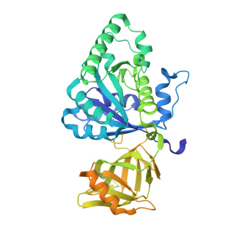
eIF5B employs a novel domain release mechanism to catalyze ribosomal subunit joining.

Kuhle, B.Ficner, R.

(2014) EMBO J 33: 1177-1191

  • DOI: https://doi.org/10.1002/embj.201387344
  • Primary Citation Related Structures: 
    4N3G, 4N3N, 4N3S, 4NCF, 4NCL, 4NCN

  • PubMed Abstract: 

    eIF5B is a eukaryal translational GTPase that catalyzes ribosomal subunit joining to form elongation-competent ribosomes. Despite its central role in protein synthesis, the mechanistic details that govern the function of eIF5B or its archaeal and bacterial (IF2) orthologs remained unclear. Here, we present six high-resolution crystal structures of eIF5B in its apo, GDP- and GTP-bound form that, together with an analysis of the thermodynamics of nucleotide binding, provide a detailed picture of the entire nucleotide cycle performed by eIF5B. Our data show that GTP binding induces significant conformational changes in the two conserved switch regions of the G domain, resulting in the reorganization of the GTPase center. These rearrangements are accompanied by the rotation of domain II relative to the G domain and release of domain III from its stable contacts with switch 2, causing an increased intrinsic flexibility in the free GTP-bound eIF5B. Based on these data, we propose a novel domain release mechanism for eIF5B/IF2 activation that explains how eIF5B and IF2 fulfill their catalytic role during ribosomal subunit joining.


  • Organizational Affiliation
    • Abteilung für Molekulare Strukturbiologie, Institut für Mikrobiologie und Genetik Göttinger Zentrum für Molekulare Biowissenschaften Georg-August-Universität Göttingen, Göttingen, Germany bkuhle@gwdg.de.

Macromolecule Content 

  • Total Structure Weight: 103.79 kDa 
  • Atom Count: 6,087 
  • Modeled Residue Count: 689 
  • Deposited Residue Count: 914 
  • Unique protein chains: 1

Macromolecules

Find similar proteins by:|  3D Structure
Entity ID: 1
MoleculeChains  Sequence LengthOrganismDetailsImage
Eukaryotic translation initiation factor 5B-like protein
A, B
457Thermochaetoides thermophila DSM 1495Mutation(s): 0 
Gene Names: CTHT_0029840
EC: 3.6.5.3
UniProt
Find proteins for G0S8G9 (Chaetomium thermophilum (strain DSM 1495 / CBS 144.50 / IMI 039719))
Explore G0S8G9 
Go to UniProtKB:  G0S8G9
Entity Groups
Sequence Clusters30% Identity50% Identity70% Identity90% Identity95% Identity100% Identity
UniProt GroupG0S8G9
Sequence Annotations
Expand
Reference Sequence

Small Molecules

Ligands 5 Unique
IDChains Name / Formula / InChI Key2D Diagram3D Interactions
GTP

Query on GTP


Download:Ideal Coordinates CCD File
C [auth A],
I [auth B]
GUANOSINE-5'-TRIPHOSPHATE
C10 H16 N5 O14 P3
XKMLYUALXHKNFT-UUOKFMHZSA-N
GOL

Query on GOL


Download:Ideal Coordinates CCD File
F [auth A]
G [auth A]
H [auth A]
M [auth B]
N [auth B]
F [auth A],
G [auth A],
H [auth A],
M [auth B],
N [auth B],
O [auth B],
P [auth B]
GLYCEROL
C3 H8 O3
PEDCQBHIVMGVHV-UHFFFAOYSA-N
ACY

Query on ACY


Download:Ideal Coordinates CCD File
L [auth B]ACETIC ACID
C2 H4 O2
QTBSBXVTEAMEQO-UHFFFAOYSA-N
MG

Query on MG


Download:Ideal Coordinates CCD File
D [auth A],
J [auth B]
MAGNESIUM ION
Mg
JLVVSXFLKOJNIY-UHFFFAOYSA-N
NA

Query on NA


Download:Ideal Coordinates CCD File
E [auth A],
K [auth B]
SODIUM ION
Na
FKNQFGJONOIPTF-UHFFFAOYSA-N

Experimental Data & Validation

Experimental Data

  • Method: X-RAY DIFFRACTION
  • Resolution: 1.87 Å
  • R-Value Free:  0.204 (Depositor), 0.205 (DCC) 
  • R-Value Work:  0.164 (Depositor), 0.166 (DCC) 
  • R-Value Observed: 0.166 (Depositor) 
Space Group: P 1 21 1
Unit Cell:
Length ( Å )Angle ( ˚ )
a = 55.41α = 90
b = 114.84β = 102.41
c = 65.88γ = 90
Software Package:
Software NamePurpose
XSCALEdata scaling
PHASERphasing
PHENIXrefinement
PDB_EXTRACTdata extraction

Structure Validation

View Full Validation Report


Ligand Structure Quality Assessment 


Entry History 

Deposition Data

Revision History  (Full details and data files)

  • Version 1.0: 2014-07-09
    Type: Initial release
  • Version 1.1: 2014-08-06
    Changes: Structure summary
  • Version 1.2: 2017-10-11
    Changes: Data collection
  • Version 1.3: 2023-09-20
    Changes: Data collection, Database references, Derived calculations, Refinement description