4N3E | pdb_00004n3e

Crystal structure of Hyp-1, a St John's wort PR-10 protein, in complex with 8-anilino-1-naphthalene sulfonate (ANS)


Experimental Data Snapshot

  • Method: X-RAY DIFFRACTION
  • Resolution: 2.43 Å
  • R-Value Free: 
    0.278 (Depositor), 0.269 (DCC) 
  • R-Value Work: 
    0.223 (Depositor), 0.225 (DCC) 
  • R-Value Observed: 
    0.224 (Depositor) 

Starting Model: experimental
View more details

wwPDB Validation 3D Report Full Report

Validation slider image for 4N3E

Ligand Structure Quality Assessment 


This is version 1.3 of the entry. See complete history

Literature

Likelihood-based molecular-replacement solution for a highly pathological crystal with tetartohedral twinning and sevenfold translational noncrystallographic symmetry.

Sliwiak, J.Jaskolski, M.Dauter, Z.McCoy, A.J.Read, R.J.

(2014) Acta Crystallogr D Biol Crystallogr 70: 471-480

  • DOI: https://doi.org/10.1107/S1399004713030319
  • Primary Citation Related Structures: 
    4N3E

  • PubMed Abstract: 

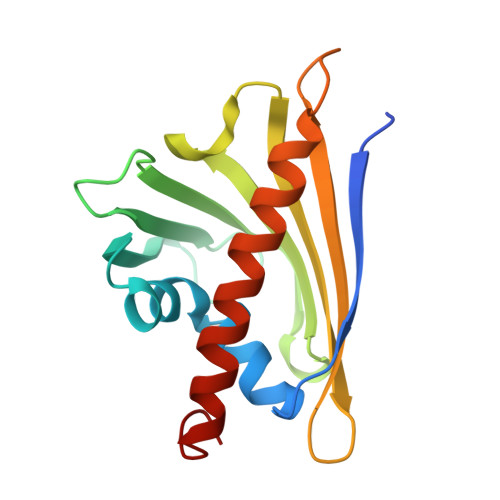
    Translational noncrystallographic symmetry (tNCS) is a pathology of protein crystals in which multiple copies of a molecule or assembly are found in similar orientations. Structure solution is problematic because this breaks the assumptions used in current likelihood-based methods. To cope with such cases, new likelihood approaches have been developed and implemented in Phaser to account for the statistical effects of tNCS in molecular replacement. Using these new approaches, it was possible to solve the crystal structure of a protein exhibiting an extreme form of this pathology with seven tetrameric assemblies arrayed along the c axis. To resolve space-group ambiguities caused by tetartohedral twinning, the structure was initially solved by placing 56 copies of the monomer in space group P1 and using the symmetry of the solution to define the true space group, C2. The resulting structure of Hyp-1, a pathogenesis-related class 10 (PR-10) protein from the medicinal herb St John's wort, reveals the binding modes of the fluorescent probe 8-anilino-1-naphthalene sulfonate (ANS), providing insight into the function of the protein in binding or storing hydrophobic ligands.


  • Organizational Affiliation
    • Center for Biocrystallographic Research, Institute of Bioorganic Chemistry, Polish Academy of Sciences, Noskowskiego 12/14, 61-704 Poznan, Poland.

Macromolecule Content 

  • Total Structure Weight: 545.27 kDa 
  • Atom Count: 37,252 
  • Modeled Residue Count: 4,452 
  • Deposited Residue Count: 4,620 
  • Unique protein chains: 1

Macromolecules

Find similar proteins by:|  3D Structure
Entity ID: 1
MoleculeChains  Sequence LengthOrganismDetailsImage
Phenolic oxidative coupling protein
A,
AA [auth a],
B,
BA [auth b],
C,
D,
E,
F,
G,
H,
I,
J,
K,
L,
M,
N,
O,
P,
Q,
R,
S,
T,
U,
V,
W,
X,
Y,
Z
165Hypericum perforatumMutation(s): 0 
Gene Names: hyp-1HYP1.1
UniProt
Find proteins for Q8H1L1 (Hypericum perforatum)
Explore Q8H1L1 
Go to UniProtKB:  Q8H1L1
Entity Groups
Sequence Clusters30% Identity50% Identity70% Identity90% Identity95% Identity100% Identity
UniProt GroupQ8H1L1
Sequence Annotations
Expand
Reference Sequence

Small Molecules

Ligands 3 Unique
IDChains Name / Formula / InChI Key2D Diagram3D Interactions
2AN

Query on 2AN


Download:Ideal Coordinates CCD File
AB [auth G]
AC [auth N]
AD [auth W]
BB [auth G]
BC [auth N]
AB [auth G],
AC [auth N],
AD [auth W],
BB [auth G],
BC [auth N],
BD [auth X],
CA [auth A],
CB [auth G],
CC [auth N],
CD [auth X],
DA [auth A],
DB [auth G],
DC [auth O],
DD [auth X],
EA [auth A],
EB [auth H],
EC [auth O],
FA [auth A],
FB [auth H],
FC [auth P],
FD [auth Y],
GA [auth B],
GB [auth H],
GC [auth P],
GD [auth Y],
HA [auth B],
HB [auth I],
HC [auth Q],
HD [auth Y],
IA [auth B],
IB [auth I],
IC [auth Q],
ID [auth Y],
JA [auth B],
JB [auth I],
JC [auth Q],
JD [auth Z],
KA [auth B],
KB [auth I],
KC [auth R],
KD [auth Z],
LA [auth C],
LB [auth I],
LC [auth R],
LD [auth Z],
MA [auth C],
MB [auth J],
MC [auth R],
NA [auth C],
NB [auth J],
NC [auth R],
ND [auth a],
OA [auth C],
OB [auth K],
OC [auth S],
OD [auth b],
PB [auth K],
PC [auth U],
PD [auth b],
QA [auth D],
QB [auth K],
QC [auth U],
QD [auth b],
RA [auth D],
RB [auth L],
RC [auth U],
SA [auth D],
SB [auth L],
SC [auth U],
TA [auth E],
TB [auth L],
TC [auth U],
UA [auth E],
UB [auth L],
UC [auth V],
VA [auth E],
VB [auth L],
VC [auth V],
WA [auth E],
WB [auth M],
XA [auth E],
XB [auth M],
XC [auth W],
YA [auth F],
YB [auth M],
YC [auth W],
ZA [auth G],
ZB [auth N],
ZC [auth W]
8-ANILINO-1-NAPHTHALENE SULFONATE
C16 H13 N O3 S
FWEOQOXTVHGIFQ-UHFFFAOYSA-N
EPE

Query on EPE


Download:Ideal Coordinates CCD File
MD [auth Z],
PA [auth C]
4-(2-HYDROXYETHYL)-1-PIPERAZINE ETHANESULFONIC ACID
C8 H18 N2 O4 S
JKMHFZQWWAIEOD-UHFFFAOYSA-N
SO4

Query on SO4


Download:Ideal Coordinates CCD File
ED [auth X],
RD [auth b],
WC [auth V]
SULFATE ION
O4 S
QAOWNCQODCNURD-UHFFFAOYSA-L

Experimental Data & Validation

Experimental Data

  • Method: X-RAY DIFFRACTION
  • Resolution: 2.43 Å
  • R-Value Free:  0.278 (Depositor), 0.269 (DCC) 
  • R-Value Work:  0.223 (Depositor), 0.225 (DCC) 
  • R-Value Observed: 0.224 (Depositor) 
Space Group: C 1 2 1
Unit Cell:
Length ( Å )Angle ( ˚ )
a = 146.29α = 90
b = 146.29β = 90
c = 298.56γ = 90
Software Package:
Software NamePurpose
HKL-2000data collection
PHASERphasing
REFMACrefinement
HKL-2000data reduction
HKL-2000data scaling

Structure Validation

View Full Validation Report


Ligand Structure Quality Assessment 


Entry History 

Deposition Data

Revision History  (Full details and data files)

  • Version 1.0: 2014-02-26
    Type: Initial release
  • Version 1.1: 2014-12-10
    Changes: Structure summary
  • Version 1.2: 2017-11-15
    Changes: Advisory, Refinement description
  • Version 1.3: 2023-09-20
    Changes: Advisory, Data collection, Database references, Derived calculations, Refinement description