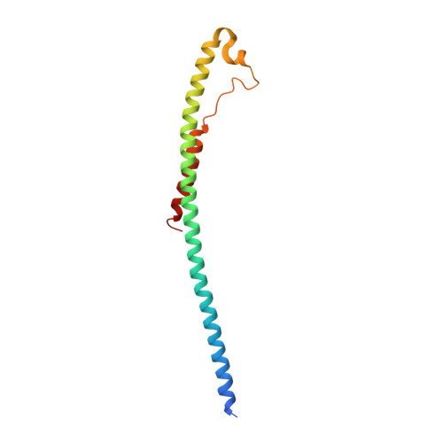
Structural Characterization of the Glycoprotein GP2 Core Domain from the CAS Virus, a Novel Arenavirus-Like Species.
Koellhoffer, J.F., Dai, Z., Malashkevich, V.N., Stenglein, M.D., Liu, Y., Toro, R., S Harrison, J., Chandran, K., Derisi, J.L., Almo, S.C., Lai, J.R.(2014) J Mol Biology 426: 1452-1468
- PubMed: 24333483
- DOI: https://doi.org/10.1016/j.jmb.2013.12.009
- Primary Citation of Related Structures:
4N21, 4N23 - PubMed Abstract:
Fusion of the viral and host cell membranes is a necessary first step for infection by enveloped viruses and is mediated by the envelope glycoprotein. The transmembrane subunits from the structurally defined "class I" glycoproteins adopt an α-helical "trimer-of-hairpins" conformation during the fusion pathway. Here, we present our studies on the envelope glycoprotein transmembrane subunit, GP2, of the CAS virus (CASV). CASV was recently identified from annulated tree boas (Corallus annulatus) with inclusion body disease and is implicated in the disease etiology. We have generated and characterized two protein constructs consisting of the predicted CASV GP2 core domain. The crystal structure of the CASV GP2 post-fusion conformation indicates a trimeric α-helical bundle that is highly similar to those of Ebola virus and Marburg virus GP2 despite CASV genome homology to arenaviruses. Denaturation studies demonstrate that the stability of CASV GP2 is pH dependent with higher stability at lower pH; we propose that this behavior is due to a network of interactions among acidic residues that would destabilize the α-helical bundle under conditions where the side chains are deprotonated. The pH-dependent stability of the post-fusion structure has been observed in Ebola virus and Marburg virus GP2, as well as other viruses that enter via the endosome. Infection experiments with CASV and the related Golden Gate virus support a mechanism of entry that requires endosomal acidification. Our results suggest that, despite being primarily arenavirus like, the transmembrane subunit of CASV is extremely similar to the filoviruses.
- Department of Biochemistry, Albert Einstein College of Medicine, 1300 Morris Park Avenue, Bronx, NY 10461, USA.
Organizational Affiliation:


