4N1U | pdb_00004n1u

Structure of human MTH1 in complex with TH588


Experimental Data Snapshot

  • Method: X-RAY DIFFRACTION
  • Resolution: 1.60 Å
  • R-Value Free: 
    0.204 (Depositor), 0.213 (DCC) 
  • R-Value Work: 
    0.180 (Depositor), 0.190 (DCC) 
  • R-Value Observed: 
    0.181 (Depositor) 

Starting Model: experimental
View more details

wwPDB Validation 3D Report Full Report

Validation slider image for 4N1U

Ligand Structure Quality Assessment 


This is version 1.4 of the entry. See complete history

Literature

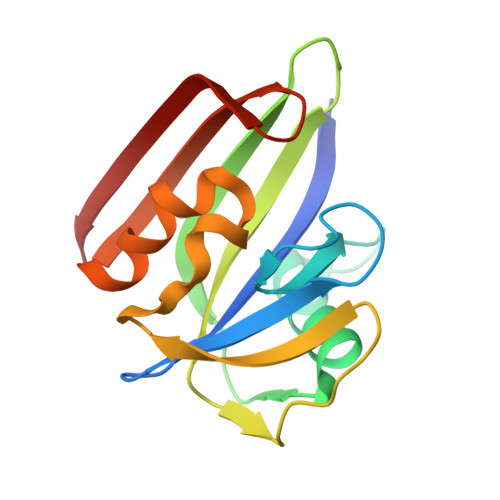
MTH1 inhibition eradicates cancer by preventing sanitation of the dNTP pool.

Gad, H.Koolmeister, T.Jemth, A.S.Eshtad, S.Jacques, S.A.Strom, C.E.Svensson, L.M.Schultz, N.Lundback, T.Einarsdottir, B.O.Saleh, A.Gokturk, C.Baranczewski, P.Svensson, R.Berntsson, R.P.Gustafsson, R.Stromberg, K.Sanjiv, K.Jacques-Cordonnier, M.C.Desroses, M.Gustavsson, A.L.Olofsson, R.Johansson, F.Homan, E.J.Loseva, O.Brautigam, L.Johansson, L.Hoglund, A.Hagenkort, A.Pham, T.Altun, M.Gaugaz, F.Z.Vikingsson, S.Evers, B.Henriksson, M.Vallin, K.S.Wallner, O.A.Hammarstrom, L.G.Wiita, E.Almlof, I.Kalderen, C.Axelsson, H.Djureinovic, T.Puigvert, J.C.Haggblad, M.Jeppsson, F.Martens, U.Lundin, C.Lundgren, B.Granelli, I.Jensen, A.J.Artursson, P.Nilsson, J.A.Stenmark, P.Scobie, M.Berglund, U.W.Helleday, T.

(2014) Nature 508: 215-221

  • DOI: https://doi.org/10.1038/nature13181
  • Primary Citation Related Structures: 
    4N1T, 4N1U

  • PubMed Abstract: 

    Cancers have dysfunctional redox regulation resulting in reactive oxygen species production, damaging both DNA and free dNTPs. The MTH1 protein sanitizes oxidized dNTP pools to prevent incorporation of damaged bases during DNA replication. Although MTH1 is non-essential in normal cells, we show that cancer cells require MTH1 activity to avoid incorporation of oxidized dNTPs, resulting in DNA damage and cell death. We validate MTH1 as an anticancer target in vivo and describe small molecules TH287 and TH588 as first-in-class nudix hydrolase family inhibitors that potently and selectively engage and inhibit the MTH1 protein in cells. Protein co-crystal structures demonstrate that the inhibitors bind in the active site of MTH1. The inhibitors cause incorporation of oxidized dNTPs in cancer cells, leading to DNA damage, cytotoxicity and therapeutic responses in patient-derived mouse xenografts. This study exemplifies the non-oncogene addiction concept for anticancer treatment and validates MTH1 as being cancer phenotypic lethal.


  • Organizational Affiliation
    • 1] Science for Life Laboratory, Division of Translational Medicine and Chemical Biology, Department of Medical Biochemistry and Biophysics, Karolinska Institutet, S-171 21 Stockholm, Sweden [2].

Macromolecule Content 

  • Total Structure Weight: 38.05 kDa 
  • Atom Count: 2,897 
  • Modeled Residue Count: 307 
  • Deposited Residue Count: 316 
  • Unique protein chains: 1

Macromolecules

Find similar proteins by:|  3D Structure
Entity ID: 1
MoleculeChains  Sequence LengthOrganismDetailsImage
7,8-dihydro-8-oxoguanine triphosphatase
A, B
158Homo sapiensMutation(s): 0 
Gene Names: NUDT1MTH1
EC: 3.6.1.55 (PDB Primary Data), 3.6.1.56 (PDB Primary Data), 3.6.1 (UniProt)
UniProt & NIH Common Fund Data Resources
Find proteins for P36639 (Homo sapiens)
Explore P36639 
Go to UniProtKB:  P36639
PHAROS:  P36639
GTEx:  ENSG00000106268 
Entity Groups
Sequence Clusters30% Identity50% Identity70% Identity90% Identity95% Identity100% Identity
UniProt GroupP36639
Sequence Annotations
Expand
Reference Sequence

Small Molecules

Ligands 2 Unique
IDChains Name / Formula / InChI Key2D Diagram3D Interactions
2GE

Query on 2GE


Download:Ideal Coordinates CCD File
C [auth A],
K [auth B]
N~4~-cyclopropyl-6-(2,3-dichlorophenyl)pyrimidine-2,4-diamine
C13 H12 Cl2 N4
PNMYJIOQIAEYQL-UHFFFAOYSA-N
SO4

Query on SO4


Download:Ideal Coordinates CCD File
D [auth A]
E [auth A]
F [auth A]
G [auth A]
H [auth A]
D [auth A],
E [auth A],
F [auth A],
G [auth A],
H [auth A],
I [auth A],
J [auth A],
L [auth B],
M [auth B],
N [auth B],
O [auth B],
P [auth B]
SULFATE ION
O4 S
QAOWNCQODCNURD-UHFFFAOYSA-L

Experimental Data & Validation

Experimental Data

  • Method: X-RAY DIFFRACTION
  • Resolution: 1.60 Å
  • R-Value Free:  0.204 (Depositor), 0.213 (DCC) 
  • R-Value Work:  0.180 (Depositor), 0.190 (DCC) 
  • R-Value Observed: 0.181 (Depositor) 
Space Group: P 21 21 21
Unit Cell:
Length ( Å )Angle ( ˚ )
a = 57.658α = 90
b = 66.073β = 90
c = 72.477γ = 90
Software Package:
Software NamePurpose
SCALAdata scaling
REFMACrefinement
PDB_EXTRACTdata extraction
XSCALEdata scaling

Structure Validation

View Full Validation Report


Ligand Structure Quality Assessment 


Entry History 

Deposition Data

Revision History  (Full details and data files)

  • Version 1.0: 2014-04-16
    Type: Initial release
  • Version 1.1: 2014-06-11
    Changes: Database references
  • Version 1.2: 2015-08-05
    Changes: Structure summary
  • Version 1.3: 2018-01-24
    Changes: Structure summary
  • Version 1.4: 2023-09-20
    Changes: Data collection, Database references, Derived calculations, Refinement description