4MZ2 | pdb_00004mz2

Crystal structure of the voltage-gated sodium channel beta 4 subunit extracellular domain


Experimental Data Snapshot

  • Method: X-RAY DIFFRACTION
  • Resolution: 1.72 Å
  • R-Value Free: 
    0.228 (Depositor), 0.230 (DCC) 
  • R-Value Work: 
    0.188 (Depositor), 0.189 (DCC) 
  • R-Value Observed: 
    0.190 (Depositor) 

Starting Model: experimental
View more details

wwPDB Validation 3D Report Full Report

Validation slider image for 4MZ2

This is version 1.3 of the entry. See complete history

Literature

Crystallographic insights into sodium-channel modulation by the beta 4 subunit.

Gilchrist, J.Das, S.Van Petegem, F.Bosmans, F.

(2013) Proc Natl Acad Sci U S A 110: E5016-E5024

  • DOI: https://doi.org/10.1073/pnas.1314557110
  • Primary Citation Related Structures: 
    4MZ2, 4MZ3

  • PubMed Abstract: 

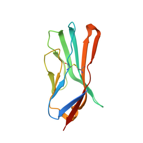
    Voltage-gated sodium (Nav) channels are embedded in a multicomponent membrane signaling complex that plays a crucial role in cellular excitability. Although the mechanism remains unclear, β-subunits modify Nav channel function and cause debilitating disorders when mutated. While investigating whether β-subunits also influence ligand interactions, we found that β4 dramatically alters toxin binding to Nav1.2. To explore these observations further, we solved the crystal structure of the extracellular β4 domain and identified (58)Cys as an exposed residue that, when mutated, eliminates the influence of β4 on toxin pharmacology. Moreover, our results suggest the presence of a docking site that is maintained by a cysteine bridge buried within the hydrophobic core of β4. Disrupting this bridge by introducing a β1 mutation implicated in epilepsy repositions the (58)Cys-containing loop and disrupts β4 modulation of Nav1.2. Overall, the principles emerging from this work (i) help explain tissue-dependent variations in Nav channel pharmacology; (ii) enable the mechanistic interpretation of β-subunit-related disorders; and (iii) provide insights in designing molecules capable of correcting aberrant β-subunit behavior.


  • Organizational Affiliation
    • Department of Physiology and Solomon H. Snyder Department of Neuroscience, The Johns Hopkins University School of Medicine, Baltimore, MD 21205.

Macromolecule Content 

  • Total Structure Weight: 14.57 kDa 
  • Atom Count: 1,038 
  • Modeled Residue Count: 111 
  • Deposited Residue Count: 129 
  • Unique protein chains: 1

Macromolecules

Find similar proteins by:|  3D Structure
Entity ID: 1
MoleculeChains  Sequence LengthOrganismDetailsImage
Sodium channel subunit beta-4129Homo sapiensMutation(s): 1 
Gene Names: SCN4B
UniProt & NIH Common Fund Data Resources
Find proteins for Q8IWT1 (Homo sapiens)
Explore Q8IWT1 
Go to UniProtKB:  Q8IWT1
PHAROS:  Q8IWT1
GTEx:  ENSG00000177098 
Entity Groups
Sequence Clusters30% Identity50% Identity70% Identity90% Identity95% Identity100% Identity
UniProt GroupQ8IWT1
Sequence Annotations
Expand
Reference Sequence

Experimental Data & Validation

Experimental Data

  • Method: X-RAY DIFFRACTION
  • Resolution: 1.72 Å
  • R-Value Free:  0.228 (Depositor), 0.230 (DCC) 
  • R-Value Work:  0.188 (Depositor), 0.189 (DCC) 
  • R-Value Observed: 0.190 (Depositor) 
Space Group: P 32 2 1
Unit Cell:
Length ( Å )Angle ( ˚ )
a = 43.445α = 90
b = 43.445β = 90
c = 108.377γ = 120
Software Package:
Software NamePurpose
XDSdata scaling
PHASERphasing
PHENIXrefinement
XDSdata reduction
XSCALEdata scaling

Structure Validation

View Full Validation Report


Entry History 

Deposition Data

Revision History  (Full details and data files)

  • Version 1.0: 2013-12-04
    Type: Initial release
  • Version 1.1: 2014-01-15
    Changes: Database references
  • Version 1.2: 2023-09-20
    Changes: Data collection, Database references, Refinement description
  • Version 1.3: 2024-10-30
    Changes: Structure summary