4MYQ | pdb_00004myq

Selective Inhibition of the Catalytic Domain Of Human Phosphodiesterase 4B With A-33


Experimental Data Snapshot

  • Method: X-RAY DIFFRACTION
  • Resolution: 1.90 Å
  • R-Value Free: 
    0.222 (Depositor), 0.224 (DCC) 
  • R-Value Work: 
    0.188 (Depositor), 0.192 (DCC) 
  • R-Value Observed: 
    0.190 (Depositor) 

Starting Model: experimental
View more details

wwPDB Validation 3D Report Full Report

Validation slider image for 4MYQ

Ligand Structure Quality Assessment 


This is version 1.4 of the entry. See complete history

Literature

Structural basis for the design of selective phosphodiesterase 4B inhibitors.

Fox, D.Burgin, A.B.Gurney, M.E.

(2014) Cell Signal 26: 657-663

  • DOI: https://doi.org/10.1016/j.cellsig.2013.12.003
  • Primary Citation Related Structures: 
    4MYQ

  • PubMed Abstract: 

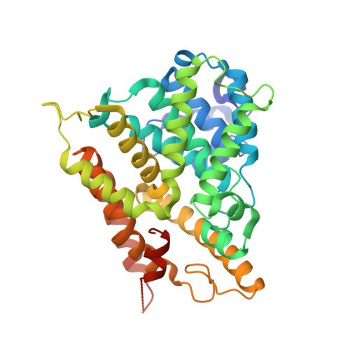
    Phosphodiesterase-4B (PDE4B) regulates the pro-inflammatory Toll Receptor -Tumor Necrosis Factor α (TNFα) pathway in monocytes, macrophages and microglial cells. As such, it is an important, although under-exploited molecular target for anti-inflammatory drugs. This is due in part to the difficulty of developing selective PDE4B inhibitors as the amino acid sequence of the PDE4 active site is identical in all PDE4 subtypes (PDE4A-D). We show that highly selective PDE4B inhibitors can be designed by exploiting sequence differences outside the active site. Specifically, PDE4B selectivity can be achieved by capture of a C-terminal regulatory helix, now termed CR3 (Control Region 3), across the active site in a conformation that closes access by cAMP. PDE4B selectivity is driven by a single amino acid polymorphism in CR3 (Leu674 in PDE4B1 versus Gln594 in PDE4D). The reciprocal mutations in PDE4B and PDE4D cause a 70-80 fold shift in selectivity. Our structural studies show that CR3 is flexible and can adopt multiple orientations and multiple registries in the closed conformation. The new co-crystal structure with bound ligand provides a guide map for the design of PDE4B selective anti-inflammatory drugs.


  • Organizational Affiliation
    • Emerald Bio, Bainbridge Island, WA, USA.

Macromolecule Content 

  • Total Structure Weight: 43.5 kDa 
  • Atom Count: 3,223 
  • Modeled Residue Count: 345 
  • Deposited Residue Count: 372 
  • Unique protein chains: 1

Macromolecules

Find similar proteins by:|  3D Structure
Entity ID: 1
MoleculeChains  Sequence LengthOrganismDetailsImage
cAMP-specific 3',5'-cyclic phosphodiesterase 4B372Homo sapiensMutation(s): 0 
Gene Names: DPDE4PDE4PDE4B
EC: 3.1.4.17 (PDB Primary Data), 3.1.4.53 (UniProt)
UniProt & NIH Common Fund Data Resources
Find proteins for Q07343 (Homo sapiens)
Explore Q07343 
Go to UniProtKB:  Q07343
PHAROS:  Q07343
GTEx:  ENSG00000184588 
Entity Groups
Sequence Clusters30% Identity50% Identity70% Identity90% Identity95% Identity100% Identity
UniProt GroupQ07343
Sequence Annotations
Expand
Reference Sequence

Small Molecules

Ligands 5 Unique
IDChains Name / Formula / InChI Key2D Diagram3D Interactions
19T

Query on 19T


Download:Ideal Coordinates CCD File
B [auth A](4-{[2-(5-chlorothiophen-2-yl)-5-ethyl-6-methylpyrimidin-4-yl]amino}phenyl)acetic acid
C19 H18 Cl N3 O2 S
FDVSPBLZPJMXFV-UHFFFAOYSA-N
ZN

Query on ZN


Download:Ideal Coordinates CCD File
D [auth A]ZINC ION
Zn
PTFCDOFLOPIGGS-UHFFFAOYSA-N
EDO

Query on EDO


Download:Ideal Coordinates CCD File
F [auth A],
G [auth A],
H [auth A]
1,2-ETHANEDIOL
C2 H6 O2
LYCAIKOWRPUZTN-UHFFFAOYSA-N
MG

Query on MG


Download:Ideal Coordinates CCD File
C [auth A]MAGNESIUM ION
Mg
JLVVSXFLKOJNIY-UHFFFAOYSA-N
NA

Query on NA


Download:Ideal Coordinates CCD File
E [auth A]SODIUM ION
Na
FKNQFGJONOIPTF-UHFFFAOYSA-N

Experimental Data & Validation

Experimental Data

  • Method: X-RAY DIFFRACTION
  • Resolution: 1.90 Å
  • R-Value Free:  0.222 (Depositor), 0.224 (DCC) 
  • R-Value Work:  0.188 (Depositor), 0.192 (DCC) 
  • R-Value Observed: 0.190 (Depositor) 
Space Group: P 42
Unit Cell:
Length ( Å )Angle ( ˚ )
a = 99.7α = 90
b = 99.7β = 90
c = 47.02γ = 90
Software Package:
Software NamePurpose
PHASERphasing
REFMACrefinement
XDSdata reduction
XSCALEdata scaling

Structure Validation

View Full Validation Report


Ligand Structure Quality Assessment 


Entry History 

Deposition Data

Revision History  (Full details and data files)

  • Version 1.0: 2014-01-01
    Type: Initial release
  • Version 1.1: 2014-01-15
    Changes: Database references
  • Version 1.2: 2014-02-05
    Changes: Database references
  • Version 1.3: 2017-11-15
    Changes: Refinement description
  • Version 1.4: 2023-09-20
    Changes: Data collection, Database references, Derived calculations, Refinement description