


Crystal Structure of Francisella tularensis 2-C-methyl-D-erythritol 4-phosphate cytidylyltransferase (IspD)
Noble, S.M., Tsang, A.K., Couch, R.D.To be published.
Experimental Data Snapshot
wwPDB Validation 3D Report Full Report

Entity ID: 1 | |||||
|---|---|---|---|---|---|
| Molecule | Chains | Sequence Length | Organism | Details | Image |
| 2-C-methyl-D-erythritol 4-phosphate cytidylyltransferase | 261 | Francisella tularensis | Mutation(s): 0 Gene Names: ispD, FTL_1525 EC: 2.7.7.60 | 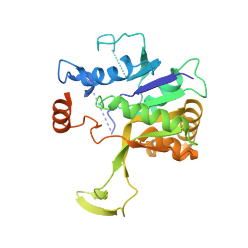 | |
| Length ( Å ) | Angle ( ˚ ) |
|---|---|
| a = 200.409 | α = 90 |
| b = 200.409 | β = 90 |
| c = 200.409 | γ = 90 |
| Software Name | Purpose |
|---|---|
| SAINT | data scaling |
| SAINT | data reduction |
| PHASER | phasing |
| PHENIX | refinement |
| PDB_EXTRACT | data extraction |
| ADSC | data collection |


