4MXC | pdb_00004mxc

Crystal structure of CMET in complex with novel inhibitor


Experimental Data Snapshot

  • Method: X-RAY DIFFRACTION
  • Resolution: 1.63 Å
  • R-Value Free: 
    0.207 (Depositor), 0.207 (DCC) 
  • R-Value Work: 
    0.179 (Depositor), 0.179 (DCC) 
  • R-Value Observed: 
    0.180 (Depositor) 

wwPDB Validation 3D Report Full Report

Validation slider image for 4MXC

Ligand Structure Quality Assessment 


This is version 1.2 of the entry. See complete history

Literature

Discovery of Anilinopyrimidines as Dual Inhibitors of c-Met and VEGFR-2: Synthesis, SAR, and Cellular Activity

Zhan, Z.S.Ai, J.Liu, Q.F.Ji, Y.C.Chen, T.T.Xu, Y.C.Geng, M.Y.Duan, W.H.

(2014) ACS Med Chem Lett 5: 673-678

  • DOI: https://doi.org/10.1021/ml500066m
  • Primary Citation Related Structures: 
    4MXC

  • PubMed Abstract: 

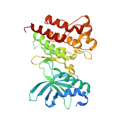
    Both c-Met and VEGFR-2 are important targets for cancer therapies. Here we report a series of potent dual c-Met and VEGFR-2 inhibitors bearing an anilinopyrimidine scaffold. Two novel synthetic protocols were employed for rapid analoguing of the designed molecules for structure-activity relationship (SAR) exploration. Some analogues displayed nanomolar potency against c-Met and VEGFR-2 at enzymatic level. Privileged compounds 3a, 3b, 3g, 3h, and 18a exhibited potent antiproliferative effect against c-Met addictive cell lines with IC50 values ranged from 0.33 to 1.7 μM. In addition, a cocrystal structure of c-Met in complex with 3h has been determined, which reveals the binding mode of c-Met to its inhibitor and helps to interpret the SAR of the analogues.


  • Organizational Affiliation
    • Department of Medicinal Chemistry, Division of Antitumor Pharmacology, and Drug Discovery and Design Center, State Key Laboratory of Drug Research, Shanghai Institute of Materia Medica, Chinese Academy of Sciences , 555 Zu Chong Zhi Road, Shanghai 201203, China.

Macromolecule Content 

  • Total Structure Weight: 36.63 kDa 
  • Atom Count: 2,570 
  • Modeled Residue Count: 290 
  • Deposited Residue Count: 319 
  • Unique protein chains: 1

Macromolecules

Find similar proteins by:|  3D Structure
Entity ID: 1
MoleculeChains  Sequence LengthOrganismDetailsImage
Hepatocyte growth factor receptor319Homo sapiensMutation(s): 0 
Gene Names: CMETMET
EC: 2.7.10.1
UniProt & NIH Common Fund Data Resources
Find proteins for P08581 (Homo sapiens)
Explore P08581 
Go to UniProtKB:  P08581
PHAROS:  P08581
GTEx:  ENSG00000105976 
Entity Groups
Sequence Clusters30% Identity50% Identity70% Identity90% Identity95% Identity100% Identity
UniProt GroupP08581
Sequence Annotations
Expand
Reference Sequence

Small Molecules

Ligands 1 Unique
IDChains Name / Formula / InChI Key2D Diagram3D Interactions
DWF

Query on DWF


Download:Ideal Coordinates CCD File
B [auth A]N-(3-fluoro-4-{[2-({3-[(methylsulfonyl)methyl]phenyl}amino)pyrimidin-4-yl]oxy}phenyl)-N'-(4-fluorophenyl)cyclopropane-1,1-dicarboxamide
C29 H25 F2 N5 O5 S
SEERCQIVLBZZJL-UHFFFAOYSA-N
Binding Affinity Annotations 
IDSourceBinding Affinity
DWF BindingDB:  4MXC IC50: 6.7 (nM) from 1 assay(s)

Experimental Data & Validation

Experimental Data

  • Method: X-RAY DIFFRACTION
  • Resolution: 1.63 Å
  • R-Value Free:  0.207 (Depositor), 0.207 (DCC) 
  • R-Value Work:  0.179 (Depositor), 0.179 (DCC) 
  • R-Value Observed: 0.180 (Depositor) 
Space Group: P 21 21 21
Unit Cell:
Length ( Å )Angle ( ˚ )
a = 42.963α = 90
b = 80.935β = 90
c = 90.867γ = 90
Software Package:
Software NamePurpose
XSCALEdata scaling
PHASERphasing
PHENIXrefinement
PDB_EXTRACTdata extraction
Blu-Icedata collection
XDSdata reduction

Structure Validation

View Full Validation Report


Ligand Structure Quality Assessment 


Entry History 

Deposition Data

Revision History  (Full details and data files)

  • Version 1.0: 2014-10-15
    Type: Initial release
  • Version 1.1: 2015-04-22
    Changes: Database references
  • Version 1.2: 2024-03-20
    Changes: Data collection, Database references, Derived calculations