4MUO | pdb_00004muo

The TrpD2 enzyme from E.coli: YbiB


Experimental Data Snapshot

  • Method: X-RAY DIFFRACTION
  • Resolution: 1.94 Å
  • R-Value Free: 
    0.212 (Depositor), 0.210 (DCC) 
  • R-Value Work: 
    0.179 (Depositor), 0.177 (DCC) 
  • R-Value Observed: 
    0.180 (Depositor) 

wwPDB Validation 3D Report Full Report

Validation slider image for 4MUO

This is version 1.3 of the entry. See complete history

Literature

YbiB from Escherichia coli, the Defining Member of the Novel TrpD2 Family of Prokaryotic DNA-binding Proteins.

Schneider, D.Kaiser, W.Stutz, C.Holinski, A.Mayans, O.Babinger, P.

(2015) J Biological Chem 290: 19527-19539

  • DOI: https://doi.org/10.1074/jbc.M114.620575
  • Primary Citation Related Structures: 
    4MUO

  • PubMed Abstract: 

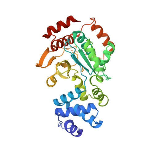
    We present the crystal structure and biochemical characterization of Escherichia coli YbiB, a member of the hitherto uncharacterized TrpD2 protein family. Our results demonstrate that the functional diversity of proteins with a common fold can be far greater than predictable by computational annotation. The TrpD2 proteins show high structural homology to anthranilate phosphoribosyltransferase (TrpD) and nucleoside phosphorylase class II enzymes but bind with high affinity (KD = 10-100 nM) to nucleic acids without detectable sequence specificity. The difference in affinity between single- and double-stranded DNA is minor. Results suggest that multiple YbiB molecules bind to one longer DNA molecule in a cooperative manner. The YbiB protein is a homodimer that, therefore, has two electropositive DNA binding grooves. But due to negative cooperativity within the dimer, only one groove binds DNA in in vitro experiments. A monomerized variant remains able to bind DNA with similar affinity, but the negative cooperative effect is eliminated. The ybiB gene forms an operon with the DNA helicase gene dinG and is under LexA control, being induced by DNA-damaging agents. Thus, speculatively, the TrpD2 proteins may be part of the LexA-controlled SOS response in bacteria.


  • Organizational Affiliation
    • From the Institute of Biophysics and Physical Biochemistry, University of Regensburg, 93040 Regensburg, Germany.

Macromolecule Content 

  • Total Structure Weight: 72.33 kDa 
  • Atom Count: 5,166 
  • Modeled Residue Count: 619 
  • Deposited Residue Count: 656 
  • Unique protein chains: 1

Macromolecules

Find similar proteins by:|  3D Structure
Entity ID: 1
MoleculeChains  Sequence LengthOrganismDetailsImage
Uncharacterized protein YbiB
A, B
328Escherichia coli K-12Mutation(s): 0 
Gene Names: ybiBb0800JW0785
UniProt
Find proteins for P30177 (Escherichia coli (strain K12))
Explore P30177 
Go to UniProtKB:  P30177
Entity Groups
Sequence Clusters30% Identity50% Identity70% Identity90% Identity95% Identity100% Identity
UniProt GroupP30177
Sequence Annotations
Expand
Reference Sequence

Experimental Data & Validation

Experimental Data

  • Method: X-RAY DIFFRACTION
  • Resolution: 1.94 Å
  • R-Value Free:  0.212 (Depositor), 0.210 (DCC) 
  • R-Value Work:  0.179 (Depositor), 0.177 (DCC) 
  • R-Value Observed: 0.180 (Depositor) 
Space Group: P 1 21 1
Unit Cell:
Length ( Å )Angle ( ˚ )
a = 45.46α = 90
b = 67.44β = 101.38
c = 105.11γ = 90
Software Package:
Software NamePurpose
SOLVEphasing
PHENIXrefinement
XDSdata reduction
XSCALEdata scaling

Structure Validation

View Full Validation Report


Entry History 

Deposition Data

Revision History  (Full details and data files)

  • Version 1.0: 2015-03-25
    Type: Initial release
  • Version 1.1: 2015-06-24
    Changes: Database references
  • Version 1.2: 2015-08-26
    Changes: Database references
  • Version 1.3: 2024-02-28
    Changes: Data collection, Database references