4MOT | pdb_00004mot

Structure of Streptococcus pneumonia pare in complex with AZ13072886


Experimental Data Snapshot

  • Method: X-RAY DIFFRACTION
  • Resolution: 1.75 Å
  • R-Value Free: 
    0.228 (Depositor), 0.236 (DCC) 
  • R-Value Work: 
    0.190 (Depositor), 0.202 (DCC) 
  • R-Value Observed: 
    0.192 (Depositor) 

wwPDB Validation 3D Report Full Report

Validation slider image for 4MOT

Ligand Structure Quality Assessment 


This is version 1.4 of the entry. See complete history

Literature

Thiazolopyridone ureas as DNA gyrase B inhibitors: Optimization of antitubercular activity and efficacy.

Kale, R.R.Kale, M.G.Waterson, D.Raichurkar, A.Hameed, S.P.Manjunatha, M.R.Kishore Reddy, B.K.Malolanarasimhan, K.Shinde, V.Koushik, K.Jena, L.K.Menasinakai, S.Humnabadkar, V.Madhavapeddi, P.Basavarajappa, H.Sharma, S.Nandishaiah, R.Mahesh Kumar, K.N.Ganguly, S.Ahuja, V.Gaonkar, S.Naveen Kumar, C.N.Ogg, D.Boriack-Sjodin, P.A.Sambandamurthy, V.K.de Sousa, S.M.Ghorpade, S.R.

(2014) Bioorg Med Chem Lett 24: 870-879

  • DOI: https://doi.org/10.1016/j.bmcl.2013.12.080
  • Primary Citation Related Structures: 
    4MOT

  • PubMed Abstract: 

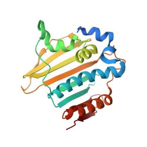
    Scaffold hopping from the thiazolopyridine ureas led to thiazolopyridone ureas with potent antitubercular activity acting through inhibition of DNA GyrB ATPase activity. Structural diversity was introduced, by extension of substituents from the thiazolopyridone N-4 position, to access hydrophobic interactions in the ribose pocket of the ATP binding region of GyrB. Further optimization of hydrogen bond interactions with arginines in site-2 of GyrB active site pocket led to potent inhibition of the enzyme (IC50 2 nM) along with potent cellular activity (MIC=0.1 μM) against Mycobacterium tuberculosis (Mtb). Efficacy was demonstrated in an acute mouse model of tuberculosis on oral administration.


  • Organizational Affiliation
    • Department of Medicinal Chemistry, AstraZeneca India Pvt. Ltd, Bellary Road, Hebbal, Bangalore 560024, India.

Macromolecule Content 

  • Total Structure Weight: 25 kDa 
  • Atom Count: 1,626 
  • Modeled Residue Count: 193 
  • Deposited Residue Count: 226 
  • Unique protein chains: 1

Macromolecules

Find similar proteins by:|  3D Structure
Entity ID: 1
MoleculeChains  Sequence LengthOrganismDetailsImage
Topoisomerase IV subunit B226Streptococcus pneumoniae R6Mutation(s): 0 
Gene Names: parEspar94_0831spr0756
EC: 5.99.1 (PDB Primary Data), 5.6.2.2 (UniProt)
UniProt
Find proteins for Q8DQB5 (Streptococcus pneumoniae (strain ATCC BAA-255 / R6))
Explore Q8DQB5 
Go to UniProtKB:  Q8DQB5
Entity Groups
Sequence Clusters30% Identity50% Identity70% Identity90% Identity95% Identity100% Identity
UniProt GroupQ8DQB5
Sequence Annotations
Expand
Reference Sequence

Small Molecules

Ligands 1 Unique
IDChains Name / Formula / InChI Key2D Diagram3D Interactions
2B7

Query on 2B7


Download:Ideal Coordinates CCD File
B [auth A]1-[4-(3-methylbutyl)-5-oxo-6-(pyridin-3-yl)-4,5-dihydro[1,3]thiazolo[5,4-b]pyridin-2-yl]-3-prop-2-en-1-ylurea
C20 H23 N5 O2 S
HDSYKPYXRVXIRI-UHFFFAOYSA-N

Experimental Data & Validation

Experimental Data

  • Method: X-RAY DIFFRACTION
  • Resolution: 1.75 Å
  • R-Value Free:  0.228 (Depositor), 0.236 (DCC) 
  • R-Value Work:  0.190 (Depositor), 0.202 (DCC) 
  • R-Value Observed: 0.192 (Depositor) 
Space Group: C 2 2 21
Unit Cell:
Length ( Å )Angle ( ˚ )
a = 73.603α = 90
b = 94.571β = 90
c = 61.001γ = 90
Software Package:
Software NamePurpose
StructureStudiodata collection
AMoREphasing
REFMACrefinement
d*TREKdata reduction
d*TREKdata scaling

Structure Validation

View Full Validation Report


Ligand Structure Quality Assessment 


Entry History 

Deposition Data

Revision History  (Full details and data files)

  • Version 1.0: 2013-11-20
    Type: Initial release
  • Version 1.1: 2013-11-27
    Changes: Data collection
  • Version 1.2: 2014-02-12
    Changes: Database references
  • Version 1.3: 2018-01-24
    Changes: Structure summary
  • Version 1.4: 2024-02-28
    Changes: Data collection, Database references, Derived calculations