4MNN | pdb_00004mnn

The crystal structure of Sso1120 from Sulfolobus solfataricus


Experimental Data Snapshot

  • Method: X-RAY DIFFRACTION
  • Resolution: 1.80 Å
  • R-Value Free: 
    0.235 (Depositor), 0.235 (DCC) 
  • R-Value Work: 
    0.193 (Depositor), 0.196 (DCC) 

Starting Model: experimental
View more details

wwPDB Validation 3D Report Full Report

Validation slider image for 4MNN

This is version 1.3 of the entry. See complete history

Literature

Sulfolobus solfataricus thiol redox puzzle: characterization of an atypical protein disulfide oxidoreductase.

Limauro, D.De Simone, G.Pirone, L.Bartolucci, S.D'Ambrosio, K.Pedone, E.

(2014) Extremophiles 18: 219-228

  • DOI: https://doi.org/10.1007/s00792-013-0607-8
  • Primary Citation Related Structures: 
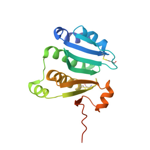
    4MNN

  • PubMed Abstract: 

    Protein disulfide oxidoreductases (PDOs) are proteins involved in disulfide bond formation playing a crucial role in adaptation to extreme environment. This paper reports the functional and structural characterization of Sso1120, a PDO from the hyperthermophilic archaeon Sulfolobus solfataricus. The protein was expressed in Escherichia coli and purified to homogeneity. The functional characterization showed that the enzyme has reductase activity, as tested by insulin assay, but differently from the other PDOs, it does not present isomerase activity. In addition it is able to form a redox couple with the thioredoxin reductase that could be used in undiscovered pathways. The protein revealed a melting point of around 90 °C in CD spectroscopy-monitored thermal denaturation and high denaturant resistance. The X-ray crystallographic structure was solved at 1.80 Å resolution, showing differences with respect to other PDOs and an unexpected similarity with the N-terminal domain of the alkyl hydroperoxide reductase F component from Salmonella typhimurium. On the basis of the reported data and of bioinformatics and phylogenetic analyses, a possible involvement of this atypical PDO in a new antioxidant system of S. solfataricus has been proposed.


  • Organizational Affiliation
    • Dipartimento di Biologia, Università degli Studi di Napoli ''Federico II'', Complesso Universitario Monte S. Angelo, Via Cinthia, 80126, Naples, Italy.

Macromolecule Content 

  • Total Structure Weight: 21.12 kDa 
  • Atom Count: 1,563 
  • Modeled Residue Count: 165 
  • Deposited Residue Count: 179 
  • Unique protein chains: 1

Macromolecules

Find similar proteins by:|  3D Structure
Entity ID: 1
MoleculeChains  Sequence LengthOrganismDetailsImage
Glutaredoxin related protein179Saccharolobus solfataricus P2Mutation(s): 0 
Gene Names: SSO1120
UniProt
Find proteins for Q97Z21 (Saccharolobus solfataricus (strain ATCC 35092 / DSM 1617 / JCM 11322 / P2))
Explore Q97Z21 
Go to UniProtKB:  Q97Z21
Entity Groups
Sequence Clusters30% Identity50% Identity70% Identity90% Identity95% Identity100% Identity
UniProt GroupQ97Z21
Sequence Annotations
Expand
Reference Sequence

Small Molecules

Ligands 1 Unique
IDChains Name / Formula / InChI Key2D Diagram3D Interactions
GOL

Query on GOL


Download:Ideal Coordinates CCD File
B [auth A]GLYCEROL
C3 H8 O3
PEDCQBHIVMGVHV-UHFFFAOYSA-N

Experimental Data & Validation

Experimental Data

  • Method: X-RAY DIFFRACTION
  • Resolution: 1.80 Å
  • R-Value Free:  0.235 (Depositor), 0.235 (DCC) 
  • R-Value Work:  0.193 (Depositor), 0.196 (DCC) 
Space Group: C 2 2 21
Unit Cell:
Length ( Å )Angle ( ˚ )
a = 68.909α = 90
b = 79.725β = 90
c = 67.65γ = 90
Software Package:
Software NamePurpose
CNSrefinement
HKL-2000data reduction
HKL-2000data scaling
CNSphasing

Structure Validation

View Full Validation Report


Entry History 

Deposition Data

Revision History  (Full details and data files)

  • Version 1.0: 2014-01-01
    Type: Initial release
  • Version 1.1: 2014-03-19
    Changes: Database references
  • Version 1.2: 2023-09-20
    Changes: Data collection, Database references, Derived calculations, Refinement description
  • Version 1.3: 2024-11-27
    Changes: Structure summary