4MHJ

Crystal structure of Fab H5M9 in complex with influenza virus hemagglutinin from A/goose/Guangdong/1/96 (H5N1)


Experimental Data Snapshot

  • Method: X-RAY DIFFRACTION
  • Resolution: 6.98 Å
  • R-Value Free: 0.387 
  • R-Value Work: 0.377 
  • R-Value Observed: 0.380 

Starting Models: experimental
View more details

wwPDB Validation   3D Report Full Report


Ligand Structure Quality Assessment 


This is version 2.2 of the entry. See complete history


Literature

A Unique and Conserved Neutralization Epitope in H5N1 Influenza Viruses Identified by an Antibody against the A/Goose/Guangdong/1/96 Hemagglutinin.

Zhu, X.Guo, Y.H.Jiang, T.Wang, Y.D.Chan, K.H.Li, X.F.Yu, W.McBride, R.Paulson, J.C.Yuen, K.Y.Qin, C.F.Che, X.Y.Wilson, I.A.

(2013) J Virol 87: 12619-12635

  • DOI: https://doi.org/10.1128/JVI.01577-13
  • Primary Citation of Related Structures:  
    4MHH, 4MHI, 4MHJ

  • PubMed Abstract: 

    Despite substantial efforts to control and contain H5N1 influenza viruses, bird flu viruses continue to spread and evolve. Neutralizing antibodies against conserved epitopes on the viral hemagglutinin (HA) could confer immunity to the diverse H5N1 virus strains and provide information for effective vaccine design. Here, we report the characterization of a broadly neutralizing murine monoclonal antibody, H5M9, to most H5N1 clades and subclades that was elicited by immunization with viral HA of A/Goose/Guangdong/1/96 (H5N1), the immediate precursor of the current dominant strains of H5N1 viruses. The crystal structures of the Fab' fragment of H5M9 in complexes with H5 HAs of A/Vietnam/1203/2004 and A/Goose/Guangdong/1/96 reveal a conserved epitope in the HA1 vestigial esterase subdomain that is some distance from the receptor binding site and partially overlaps antigenic site C of H3 HA. Further epitope characterization by selection of escape mutants and epitope mapping by flow cytometry analysis of site-directed mutagenesis of HA with a yeast cell surface display identified four residues that are critical for H5M9 binding. D53, Y274, E83a, and N276 are all conserved in H5N1 HAs and are not in H5 epitopes identified by other mouse or human antibodies. Antibody H5M9 is effective in protection of H5N1 virus both prophylactically and therapeutically and appears to neutralize by blocking both virus receptor binding and postattachment steps. Thus, the H5M9 epitope identified here should provide valuable insights into H5N1 vaccine design and improvement, as well as antibody-based therapies for treatment of H5N1 infection.


  • Organizational Affiliation

    Department of Integrative Structural and Computational Biology, The Scripps Research Institute, La Jolla, California, USA.


Macromolecules
Find similar proteins by:  (by identity cutoff)  |  3D Structure
Entity ID: 1
MoleculeChains Sequence LengthOrganismDetailsImage
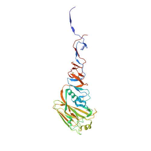
Hemagglutinin HA1 chain334Influenza A virus (A/goose/Guangdong/1/1996(H5N1))Mutation(s): 0 
Gene Names: HA
UniProt
Find proteins for Q9Q0U6 (Influenza A virus (strain A/Goose/Guangdong/1/1996 H5N1 genotype Gs/Gd))
Explore Q9Q0U6 
Go to UniProtKB:  Q9Q0U6
Entity Groups  
Sequence Clusters30% Identity50% Identity70% Identity90% Identity95% Identity100% Identity
UniProt GroupQ9Q0U6
Glycosylation
Glycosylation Sites: 2
Sequence Annotations
Expand
  • Reference Sequence
Find similar proteins by:  (by identity cutoff)  |  3D Structure
Entity ID: 2
MoleculeChains Sequence LengthOrganismDetailsImage
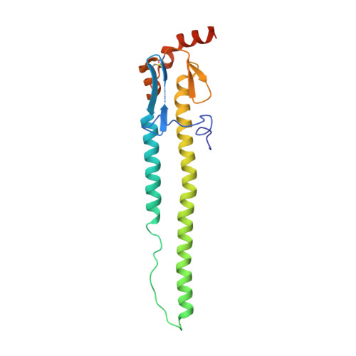
Hemagglutinin HA2 chain182Influenza A virus (A/goose/Guangdong/1/1996(H5N1))Mutation(s): 0 
Gene Names: HA
UniProt
Find proteins for Q9Q0U6 (Influenza A virus (strain A/Goose/Guangdong/1/1996 H5N1 genotype Gs/Gd))
Explore Q9Q0U6 
Go to UniProtKB:  Q9Q0U6
Entity Groups  
Sequence Clusters30% Identity50% Identity70% Identity90% Identity95% Identity100% Identity
UniProt GroupQ9Q0U6
Glycosylation
Glycosylation Sites: 1
Sequence Annotations
Expand
  • Reference Sequence
Find similar proteins by:  (by identity cutoff)  |  3D Structure
Entity ID: 3
MoleculeChains Sequence LengthOrganismDetailsImage
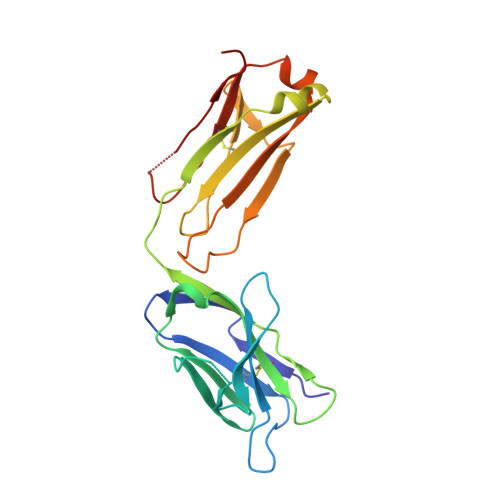
H5M9 antibody, light chain (kappa)218Mus musculusMutation(s): 0 
Entity Groups  
Sequence Clusters30% Identity50% Identity70% Identity90% Identity95% Identity100% Identity
Sequence Annotations
Expand
  • Reference Sequence
Find similar proteins by:  (by identity cutoff)  |  3D Structure
Entity ID: 4
MoleculeChains Sequence LengthOrganismDetailsImage
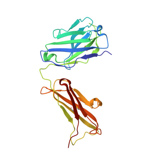
H5M9 antibody, heavy chain (IgG1)222Mus musculusMutation(s): 0 
Entity Groups  
Sequence Clusters30% Identity50% Identity70% Identity90% Identity95% Identity100% Identity
Sequence Annotations
Expand
  • Reference Sequence
Oligosaccharides

Help

Entity ID: 5
MoleculeChains Length2D Diagram Glycosylation3D Interactions
alpha-D-mannopyranose-(1-4)-2-acetamido-2-deoxy-beta-D-glucopyranose-(1-4)-2-acetamido-2-deoxy-beta-D-glucopyranoseDA [auth d],
FA [auth f],
Y
3N-Glycosylation
Glycosylation Resources
GlyTouCan:  G62182OO
GlyCosmos:  G62182OO
GlyGen:  G62182OO
Entity ID: 6
MoleculeChains Length2D Diagram Glycosylation3D Interactions
alpha-D-mannopyranose-(1-2)-alpha-D-mannopyranose-(1-3)-[alpha-D-mannopyranose-(1-2)-alpha-D-mannopyranose-(1-6)]alpha-D-mannopyranose-(1-6)-beta-D-mannopyranose-(1-4)-2-acetamido-2-deoxy-beta-D-glucopyranose-(1-4)-2-acetamido-2-deoxy-beta-D-glucopyranose
Z
8N-Glycosylation
Glycosylation Resources
GlyTouCan:  G48018RF
GlyCosmos:  G48018RF
GlyGen:  G48018RF
Entity ID: 7
MoleculeChains Length2D Diagram Glycosylation3D Interactions
2-acetamido-2-deoxy-beta-D-glucopyranose-(1-4)-2-acetamido-2-deoxy-beta-D-glucopyranoseAA [auth a],
CA [auth c]
2N-Glycosylation
Glycosylation Resources
GlyTouCan:  G42666HT
GlyCosmos:  G42666HT
GlyGen:  G42666HT
Entity ID: 8
MoleculeChains Length2D Diagram Glycosylation3D Interactions
beta-D-mannopyranose-(1-4)-2-acetamido-2-deoxy-beta-D-glucopyranose-(1-4)-2-acetamido-2-deoxy-beta-D-glucopyranoseBA [auth b]3N-Glycosylation
Glycosylation Resources
GlyTouCan:  G15407YE
GlyCosmos:  G15407YE
GlyGen:  G15407YE
Entity ID: 9
MoleculeChains Length2D Diagram Glycosylation3D Interactions
alpha-D-mannopyranose-(1-3)-beta-D-mannopyranose-(1-4)-2-acetamido-2-deoxy-beta-D-glucopyranose-(1-4)-2-acetamido-2-deoxy-beta-D-glucopyranoseEA [auth e]4N-Glycosylation
Glycosylation Resources
GlyTouCan:  G81315DD
GlyCosmos:  G81315DD
GlyGen:  G81315DD
Experimental Data & Validation

Experimental Data

  • Method: X-RAY DIFFRACTION
  • Resolution: 6.98 Å
  • R-Value Free: 0.387 
  • R-Value Work: 0.377 
  • R-Value Observed: 0.380 
  • Space Group: P 31 2 1
Unit Cell:
Length ( Å )Angle ( ˚ )
a = 199.552α = 90
b = 199.552β = 90
c = 466.886γ = 120
Software Package:
Software NamePurpose
HKL-2000data collection
PHASERphasing
REFMACrefinement
HKL-2000data reduction
HKL-2000data scaling

Structure Validation

View Full Validation Report


Ligand Structure Quality Assessment 


Entry History 

Deposition Data

Revision History  (Full details and data files)

  • Version 1.0: 2013-10-02
    Type: Initial release
  • Version 1.1: 2013-11-20
    Changes: Database references
  • Version 2.0: 2020-07-29
    Type: Remediation
    Reason: Carbohydrate remediation
    Changes: Advisory, Atomic model, Data collection, Database references, Derived calculations, Structure summary
  • Version 2.1: 2023-09-20
    Changes: Data collection, Database references, Refinement description, Structure summary
  • Version 2.2: 2024-10-09
    Changes: Structure summary