4M8M | pdb_00004m8m

Crystal structure of the active dimer of zebrafish PlexinC1 cytoplasmic region


Experimental Data Snapshot

  • Method: X-RAY DIFFRACTION
  • Resolution: 3.31 Å
  • R-Value Free: 
    0.281 (Depositor), 0.286 (DCC) 
  • R-Value Work: 
    0.226 (Depositor), 0.226 (DCC) 
  • R-Value Observed: 
    0.228 (Depositor) 

wwPDB Validation 3D Report Full Report

Validation slider image for 4M8M

This is version 1.2 of the entry. See complete history

Literature

Structural basis for activation and non-canonical catalysis of the Rap GTPase activating protein domain of plexin.

Wang, Y.Pascoe, H.G.Brautigam, C.A.He, H.Zhang, X.

(2013) Elife 2: e01279-e01279

  • DOI: https://doi.org/10.7554/eLife.01279
  • Primary Citation Related Structures: 
    4M8M, 4M8N

  • PubMed Abstract: 

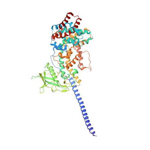
    Plexins are cell surface receptors that bind semaphorins and transduce signals for regulating neuronal axon guidance and other processes. Plexin signaling depends on their cytoplasmic GTPase activating protein (GAP) domain, which specifically inactivates the Ras homolog Rap through an ill-defined non-canonical catalytic mechanism. The plexin GAP is activated by semaphorin-induced dimerization, the structural basis for which remained unknown. Here we present the crystal structures of the active dimer of zebrafish PlexinC1 cytoplasmic region in the apo state and in complex with Rap. The structures show that the dimerization induces a large-scale conformational change in plexin, which opens the GAP active site to allow Rap binding. Plexin stabilizes the switch II region of Rap in an unprecedented conformation, bringing Gln63 in Rap into the active site for catalyzing GTP hydrolysis. The structures also explain the unique Rap-specificity of plexins. Mutational analyses support that these mechanisms underlie plexin activation and signaling. DOI:http://dx.doi.org/10.7554/eLife.01279.001.


  • Organizational Affiliation
    • Department of Pharmacology , University of Texas Southwestern Medical Center , Dallas , United States.

Macromolecule Content 

  • Total Structure Weight: 146.25 kDa 
  • Atom Count: 8,888 
  • Modeled Residue Count: 1,147 
  • Deposited Residue Count: 1,264 
  • Unique protein chains: 1

Macromolecules

Find similar proteins by:|  3D Structure
Entity ID: 1
MoleculeChains  Sequence LengthOrganismDetailsImage
GCN4 coiled-coil fused zebrafish PlexinC1
A, B
632Danio rerioMutation(s): 0 
Gene Names: Plexin C1plxnc1
UniProt
Find proteins for Q5RGW1 (Danio rerio)
Explore Q5RGW1 
Go to UniProtKB:  Q5RGW1
Entity Groups
Sequence Clusters30% Identity50% Identity70% Identity90% Identity95% Identity100% Identity
UniProt GroupQ5RGW1
Sequence Annotations
Expand
Reference Sequence

Experimental Data & Validation

Experimental Data

  • Method: X-RAY DIFFRACTION
  • Resolution: 3.31 Å
  • R-Value Free:  0.281 (Depositor), 0.286 (DCC) 
  • R-Value Work:  0.226 (Depositor), 0.226 (DCC) 
  • R-Value Observed: 0.228 (Depositor) 
Space Group: P 21 21 21
Unit Cell:
Length ( Å )Angle ( ˚ )
a = 53.216α = 90
b = 146.1β = 90
c = 209.581γ = 90
Software Package:
Software NamePurpose
HKL-3000data collection
PHASERphasing
PHENIXrefinement
HKL-3000data reduction
HKL-3000data scaling

Structure Validation

View Full Validation Report


Entry History 

Deposition Data

Revision History  (Full details and data files)

  • Version 1.0: 2013-10-09
    Type: Initial release
  • Version 1.1: 2014-01-15
    Changes: Database references
  • Version 1.2: 2024-02-28
    Changes: Data collection, Database references