


crystal structure of anti-IL-23 antibody CNTO1959 at pH 6.5
Teplyakov, A., Obmolova, G., Gilliland, G.L.To be published.
Experimental Data Snapshot
Starting Model: experimental
View more details
wwPDB Validation 3D Report Full Report

Entity ID: 1 | |||||
|---|---|---|---|---|---|
| Molecule | Chains | Sequence Length | Organism | Details | Image |
| CNTO1959 light chain | A [auth L] | 217 | Homo sapiens | Mutation(s): 0 | 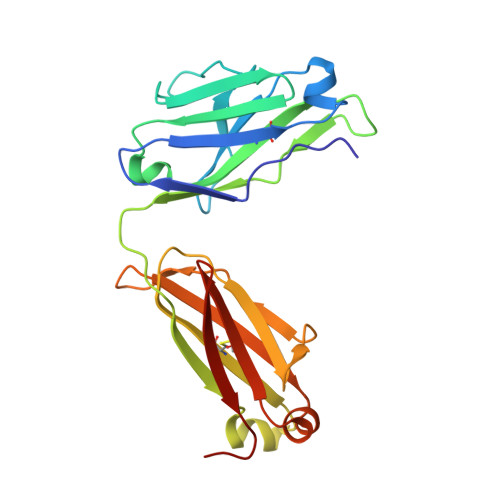 |
Entity ID: 2 | |||||
|---|---|---|---|---|---|
| Molecule | Chains | Sequence Length | Organism | Details | Image |
| CNTO1959 heavy chain | B [auth H] | 226 | Homo sapiens | Mutation(s): 0 | 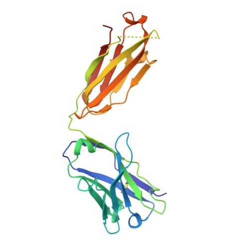 |
| Ligands 1 Unique | |||||
|---|---|---|---|---|---|
| ID | Chains | Name / Formula / InChI Key | 2D Diagram | 3D Interactions | |
| FMT Download:Ideal Coordinates CCD File | C [auth H], D [auth H] | FORMIC ACID C H2 O2 BDAGIHXWWSANSR-UHFFFAOYSA-N |  | ||
| Length ( Å ) | Angle ( ˚ ) |
|---|---|
| a = 62.59 | α = 90 |
| b = 92.05 | β = 90 |
| c = 92.82 | γ = 90 |
| Software Name | Purpose |
|---|---|
| StructureStudio | data collection |
| PHASER | phasing |
| REFMAC | refinement |
| XDS | data reduction |
| XDS | data scaling |


