4M62 | pdb_00004m62

Ontogeny of recognition specificity and functionality for the anti-HIV neutralizing antibody 4E10


Experimental Data Snapshot

  • Method: X-RAY DIFFRACTION
  • Resolution: 1.80 Å
  • R-Value Free: 
    0.216 (Depositor), 0.227 (DCC) 
  • R-Value Work: 
    0.189 (Depositor), 0.198 (DCC) 
  • R-Value Observed: 
    0.191 (Depositor) 

wwPDB Validation 3D Report Full Report

Validation slider image for 4M62

This is version 1.1 of the entry. See complete history

Literature

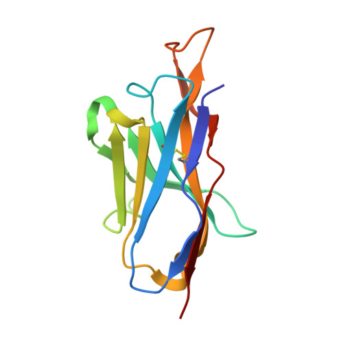
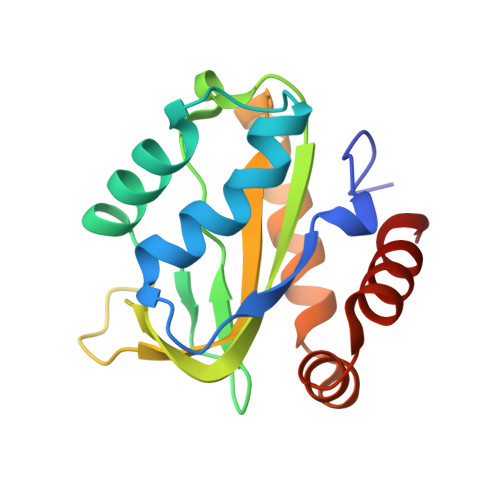
Ontogeny of Recognition Specificity and Functionality for the Broadly Neutralizing Anti-HIV Antibody 4E10.

Finton, K.A.Friend, D.Jaffe, J.Gewe, M.Holmes, M.A.Larman, H.B.Stuart, A.Larimore, K.Greenberg, P.D.Elledge, S.J.Stamatatos, L.Strong, R.K.

(2014) PLoS Pathog 10: e1004403-e1004403

  • DOI: https://doi.org/10.1371/journal.ppat.1004403
  • Primary Citation Related Structures: 
    4LRN, 4M62, 4M8Q, 4OB5, 4ODX

  • PubMed Abstract: 

    The process of antibody ontogeny typically improves affinity, on-rate, and thermostability, narrows polyspecificity, and rigidifies the combining site to the conformer optimal for binding from the broader ensemble accessible to the precursor. However, many broadly-neutralizing anti-HIV antibodies incorporate unusual structural elements and recognition specificities or properties that often lead to autoreactivity. The ontogeny of 4E10, an autoreactive antibody with unexpected combining site flexibility, was delineated through structural and biophysical comparisons of the mature antibody with multiple potential precursors. 4E10 gained affinity primarily by off-rate enhancement through a small number of mutations to a highly conserved recognition surface. Controverting the conventional paradigm, the combining site gained flexibility and autoreactivity during ontogeny, while losing thermostability, though polyspecificity was unaffected. Details of the recognition mechanism, including inferred global effects due to 4E10 binding, suggest that neutralization by 4E10 may involve mechanisms beyond simply binding, also requiring the ability of the antibody to induce conformational changes distant from its binding site. 4E10 is, therefore, unlikely to be re-elicited by conventional vaccination strategies.


  • Organizational Affiliation
    • Division of Basic Sciences, Fred Hutchinson Cancer Research Center, Seattle, Washington, United States of America.

Macromolecule Content 

  • Total Structure Weight: 87.35 kDa 
  • Atom Count: 6,793 
  • Modeled Residue Count: 789 
  • Deposited Residue Count: 790 
  • Unique protein chains: 3

Macromolecules

Find similar proteins by:|  3D Structure
Entity ID: 1
MoleculeChains  Sequence LengthOrganismDetailsImage
GEP2 FV light chainA [auth L],
C [auth M]
113Homo sapiensMutation(s): 0 
UniProt & NIH Common Fund Data Resources
Find proteins for P01619 (Homo sapiens)
Explore P01619 
Go to UniProtKB:  P01619
PHAROS:  P01619
GTEx:  ENSG00000239951 
Entity Groups
Sequence Clusters30% Identity50% Identity70% Identity90% Identity95% Identity100% Identity
UniProt GroupP01619
Sequence Annotations
Expand
Reference Sequence
Find similar proteins by:|  3D Structure
Entity ID: 2
MoleculeChains  Sequence LengthOrganismDetailsImage
GEP2 FV heavy chainB [auth H],
D [auth I]
127Homo sapiensMutation(s): 0 
Find similar proteins by:|  3D Structure
Entity ID: 3
MoleculeChains  Sequence LengthOrganismDetailsImage
T117E [auth S],
F [auth T]
155synthetic constructMutation(s): 0 

Small Molecules

Ligands 1 Unique
IDChains Name / Formula / InChI Key2D Diagram3D Interactions
SO4

Query on SO4


Download:Ideal Coordinates CCD File
G [auth L]
H [auth L]
I [auth M]
J [auth S]
K [auth S]
G [auth L],
H [auth L],
I [auth M],
J [auth S],
K [auth S],
L [auth T],
M [auth T],
N [auth T],
O [auth T]
SULFATE ION
O4 S
QAOWNCQODCNURD-UHFFFAOYSA-L

Experimental Data & Validation

Experimental Data

  • Method: X-RAY DIFFRACTION
  • Resolution: 1.80 Å
  • R-Value Free:  0.216 (Depositor), 0.227 (DCC) 
  • R-Value Work:  0.189 (Depositor), 0.198 (DCC) 
  • R-Value Observed: 0.191 (Depositor) 
Space Group: P 21 21 21
Unit Cell:
Length ( Å )Angle ( ˚ )
a = 69.88α = 90
b = 103.48β = 90
c = 109.3γ = 90
Software Package:
Software NamePurpose
CrystalCleardata collection
PHASERphasing
REFMACrefinement
d*TREKdata reduction
d*TREKdata scaling

Structure Validation

View Full Validation Report


Entry History 

Deposition Data

Revision History  (Full details and data files)

  • Version 1.0: 2014-10-08
    Type: Initial release
  • Version 1.1: 2024-11-20
    Changes: Data collection, Database references, Derived calculations, Structure summary