4M5P | pdb_00004m5p

OYE2.6 Y78W, I113C


Experimental Data Snapshot

  • Method: X-RAY DIFFRACTION
  • Resolution: 1.50 Å
  • R-Value Free: 
    0.170 (Depositor), 0.169 (DCC) 
  • R-Value Work: 
    0.141 (Depositor), 0.141 (DCC) 
  • R-Value Observed: 
    0.144 (Depositor) 

Starting Model: experimental
View more details

wwPDB Validation 3D Report Full Report

Validation slider image for 4M5P

Ligand Structure Quality Assessment 


This is version 1.4 of the entry. See complete history

Literature

Residues Controlling Facial Selectivity in an Alkene Reductase and Semirational Alterations to Create Stereocomplementary Variants.

Walton, A.Z.Sullivan, B.Patterson-Orazem, A.C.Stewart, J.D.

(2014) ACS Catal 4: 2307-2318

  • DOI: https://doi.org/10.1021/cs500429k
  • Primary Citation Related Structures: 
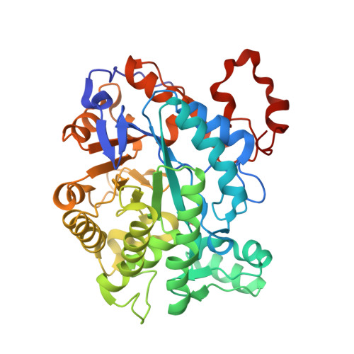
    4M5P, 4QAI

  • PubMed Abstract: 

    A systematic saturation mutagenesis campaign was carried out on an alkene reductase from Pichia stipitis (OYE 2.6) to develop variants with reversed stereoselectivities. Wild-type OYE 2.6 reduces three representative Baylis-Hillman adducts to the corresponding S products with almost complete stereoselectivities and good catalytic efficiencies. We created and screened 13 first-generation, site-saturation mutagenesis libraries, targeting residues found near the bound substrate. One variant (Tyr78Trp) showed high R selectivity toward one of the three substrates, but no change (cyclohexenone derivative) and no catalytic activity (acrylate derivative) for the other two. Subsequent rounds of mutagenesis retained the Tyr78Trp mutation and explored other residues that impacted stereoselectivity when altered in a wild-type background. These efforts yielded double and triple mutants that possessed inverted stereoselectivities for two of the three substrates (conversions >99% and at least 91% ee ( R )). To understand the reasons underlying the stereochemical changes, we solved crystal structures of two key mutants: Tyr78Trp and Tyr78Trp/Ile113Cys, the latter with substrate partially occupying the active site. By combining these experimental data with modeling studies, we have proposed a rationale that explains the impacts of the most useful mutations.


  • Organizational Affiliation
    • Department of Chemistry, 126 Sisler Hall, University of Florida , Gainesville, Florida 32611 United States.

Macromolecule Content 

  • Total Structure Weight: 46.29 kDa 
  • Atom Count: 3,852 
  • Modeled Residue Count: 405 
  • Deposited Residue Count: 408 
  • Unique protein chains: 1

Macromolecules

Find similar proteins by:|  3D Structure
Entity ID: 1
MoleculeChains  Sequence LengthOrganismDetailsImage
NADPH dehydrogenase407Scheffersomyces stipitis CBS 6054Mutation(s): 3 
Gene Names: OYE2.6PICST_44614
UniProt
Find proteins for A3LT82 (Scheffersomyces stipitis (strain ATCC 58785 / CBS 6054 / NBRC 10063 / NRRL Y-11545))
Explore A3LT82 
Go to UniProtKB:  A3LT82
Entity Groups
Sequence Clusters30% Identity50% Identity70% Identity90% Identity95% Identity100% Identity
UniProt GroupA3LT82
Sequence Annotations
Expand
Reference Sequence

Small Molecules

Ligands 4 Unique
IDChains Name / Formula / InChI Key2D Diagram3D Interactions
FMN

Query on FMN


Download:Ideal Coordinates CCD File
B [auth A]FLAVIN MONONUCLEOTIDE
C17 H21 N4 O9 P
FVTCRASFADXXNN-SCRDCRAPSA-N
23W

Query on 23W


Download:Ideal Coordinates CCD File
I [auth A]methyl 2-(hydroxymethyl)prop-2-enoate
C5 H8 O3
RFUCOAQWQVDBEU-UHFFFAOYSA-N
MLA

Query on MLA


Download:Ideal Coordinates CCD File
C [auth A],
D [auth A],
H [auth A]
MALONIC ACID
C3 H4 O4
OFOBLEOULBTSOW-UHFFFAOYSA-N
NA

Query on NA


Download:Ideal Coordinates CCD File
E [auth A],
F [auth A],
G [auth A]
SODIUM ION
Na
FKNQFGJONOIPTF-UHFFFAOYSA-N
Modified Residues  1 Unique
IDChains TypeFormula2D DiagramParent
CSD
Query on CSD
A
L-PEPTIDE LINKINGC3 H7 N O4 SCYS

Experimental Data & Validation

Experimental Data

  • Method: X-RAY DIFFRACTION
  • Resolution: 1.50 Å
  • R-Value Free:  0.170 (Depositor), 0.169 (DCC) 
  • R-Value Work:  0.141 (Depositor), 0.141 (DCC) 
  • R-Value Observed: 0.144 (Depositor) 
Space Group: P 63 2 2
Unit Cell:
Length ( Å )Angle ( ˚ )
a = 126.487α = 90
b = 126.487β = 90
c = 123.307γ = 120
Software Package:
Software NamePurpose
CBASSdata collection
AMoREphasing
PHENIXrefinement
MOSFLMdata reduction
SCALEPACKdata scaling

Structure Validation

View Full Validation Report


Ligand Structure Quality Assessment 


Entry History 

Deposition Data

Revision History  (Full details and data files)

  • Version 1.0: 2014-06-25
    Type: Initial release
  • Version 1.1: 2014-09-24
    Changes: Database references
  • Version 1.2: 2015-04-29
    Changes: Non-polymer description
  • Version 1.3: 2023-09-20
    Changes: Data collection, Database references, Derived calculations, Refinement description
  • Version 1.4: 2024-10-16
    Changes: Structure summary