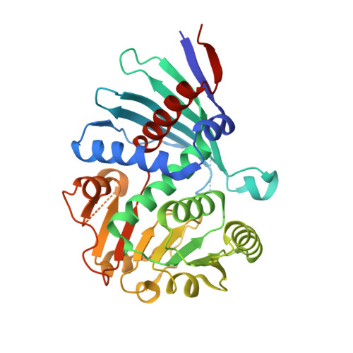
4M54 | pdb_00004m54

The structure of the staphyloferrin B precursor biosynthetic enzyme SbnB bound to N-(1-amino-1-carboxyl-2-ethyl)-glutamic acid and NADH


Experimental Data Snapshot

  • Method: X-RAY DIFFRACTION
  • Resolution: 2.36 Å
  • R-Value Free: 
    0.258 (Depositor), 0.260 (DCC) 
  • R-Value Work: 
    0.223 (Depositor), 0.220 (DCC) 
  • R-Value Observed: 
    0.227 (Depositor) 

wwPDB Validation   3D Report Full Report


Ligand Structure Quality Assessment 

Created with Raphaël 2.3.0Worse 01 BetterLigand structure goodness of fit to experimental dataBest fitted NAIClick on this verticalbar to view detailsBest fitted AE5Click on this verticalbar to view details

This is version 1.2 of the entry. See complete history


Literature

Synthesis of L-2,3-diaminopropionic acid, a siderophore and antibiotic precursor.

Kobylarz, M.J.Grigg, J.C.Takayama, S.J.Rai, D.K.Heinrichs, D.E.Murphy, M.E.

(2014) Chem Biol 21: 379-388

  • DOI: https://doi.org/10.1016/j.chembiol.2013.12.011
  • Primary Citation of Related Structures:  
    4M54, 4MP3, 4MP6, 4MP8, 4MPD

  • PubMed Abstract: 

    L-2,3-diaminopropionic acid (L-Dap) is an amino acid that is a precursor of antibiotics and staphyloferrin B a siderophore produced by Staphylococcus aureus. SbnA and SbnB are encoded by the staphyloferrin B biosynthetic gene cluster and are implicated in L-Dap biosynthesis. We demonstrate here that SbnA uses PLP and substrates O-phospho-L-serine and L-glutamate to produce a metabolite N-(1-amino-1-carboxyl-2-ethyl)-glutamic acid (ACEGA). SbnB is shown to use NAD(+) to oxidatively hydrolyze ACEGA to yield α-ketoglutarate and L-Dap. Also, we describe crystal structures of SbnB in complex with NADH and ACEGA as well as with NAD(+) and α-ketoglutarate to reveal the residues required for substrate binding, oxidation, and hydrolysis. SbnA and SbnB contribute to the iron sparing response of S. aureus that enables staphyloferrin B biosynthesis in the absence of an active tricarboxylic acid cycle.


  • Organizational Affiliation

    Department of Microbiology and Immunology, Life Sciences Institute, The University of British Columbia, Vancouver, BC V6T 1Z3, Canada.


Macromolecules
Find similar proteins by:  (by identity cutoff)  |  3D Structure
Entity ID: 1
MoleculeChains Sequence LengthOrganismDetailsImage
Putative ornithine cyclodeaminase339Staphylococcus aureus subsp. aureus MW2Mutation(s): 0 
Gene Names: sbnB
UniProt
Find proteins for A0A0H3K9Y6 (Staphylococcus aureus (strain Newman))
Explore A0A0H3K9Y6 
Go to UniProtKB:  A0A0H3K9Y6
Entity Groups  
Sequence Clusters30% Identity50% Identity70% Identity90% Identity95% Identity100% Identity
UniProt GroupA0A0H3K9Y6
Sequence Annotations
Expand
  • Reference Sequence
Experimental Data & Validation

Experimental Data

  • Method: X-RAY DIFFRACTION
  • Resolution: 2.36 Å
  • R-Value Free:  0.258 (Depositor), 0.260 (DCC) 
  • R-Value Work:  0.223 (Depositor), 0.220 (DCC) 
  • R-Value Observed: 0.227 (Depositor) 
Space Group: P 41 21 2
Unit Cell:
Length ( Å )Angle ( ˚ )
a = 63.116α = 90
b = 63.116β = 90
c = 159.413γ = 90
Software Package:
Software NamePurpose
DENZOdata reduction
SCALEPACKdata scaling
PHENIXrefinement
PDB_EXTRACTdata extraction
HKL-3000data collection
HKL-3000data reduction
HKL-3000data scaling
PHENIXphasing

Structure Validation

View Full Validation Report


Ligand Structure Quality Assessment 

Created with Raphaël 2.3.0Worse 01 BetterLigand structure goodness of fit to experimental dataBest fitted NAIClick on this verticalbar to view detailsBest fitted AE5Click on this verticalbar to view details

Entry History 

Deposition Data

Revision History  (Full details and data files)

  • Version 1.0: 2014-07-09
    Type: Initial release
  • Version 1.1: 2017-11-15
    Changes: Refinement description
  • Version 1.2: 2024-02-28
    Changes: Data collection, Database references, Derived calculations