4M4B

Human Hemoglobin Nitroethane Modified


Experimental Data Snapshot

  • Method: X-RAY DIFFRACTION
  • Resolution: 2.00 Å
  • R-Value Free: 0.270 
  • R-Value Work: 0.229 
  • R-Value Observed: 0.231 

Starting Model: experimental
View more details

wwPDB Validation   3D Report Full Report


Ligand Structure Quality Assessment 


This is version 1.1 of the entry. See complete history


Literature

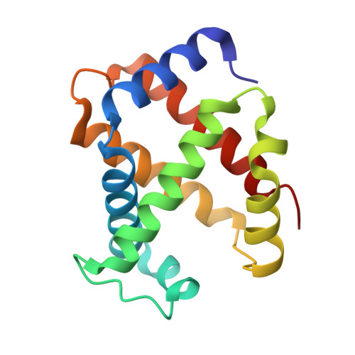
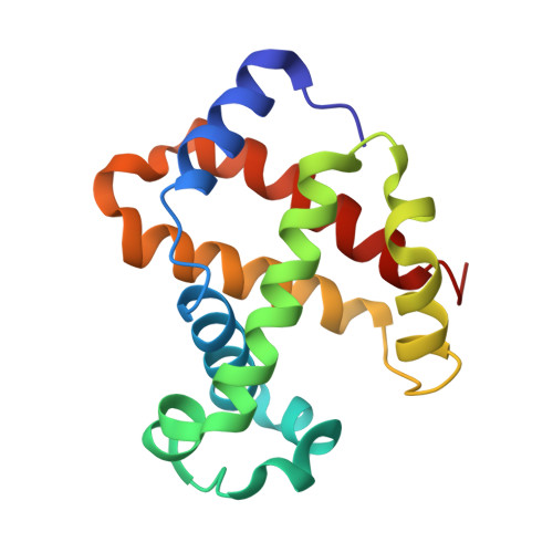
Degradation of human hemoglobin by organic C-nitroso compounds.

Yi, J.Ye, G.Thomas, L.M.Richter-Addo, G.B.

(2013) Chem Commun (Camb) 49: 11179-11181

  • DOI: https://doi.org/10.1039/c3cc46174b
  • Primary Citation of Related Structures:  
    4M4A, 4M4B

  • PubMed Abstract: 

    The crystal structure of the nitrosomethane adduct of human Hb shows N-binding of the MeNO ligands to heme Fe. The structure of the EtNO adduct reveals a surprising 4.9 Å heme slippage in the β subunit, and explains the ability of C-nitroso compounds to degrade Hb removing it from circulation.


  • Organizational Affiliation

    Department of Chemistry and Biochemistry, University of Oklahoma, Norman, Oklahoma, USA. yijun@ou.edu grichteraddo@ou.edu.


Macromolecules
Find similar proteins by:  (by identity cutoff)  |  3D Structure
Entity ID: 1
MoleculeChains Sequence LengthOrganismDetailsImage
Hemoglobin subunit alpha141Homo sapiensMutation(s): 0 
UniProt & NIH Common Fund Data Resources
Find proteins for P69905 (Homo sapiens)
Explore P69905 
Go to UniProtKB:  P69905
Entity Groups  
Sequence Clusters30% Identity50% Identity70% Identity90% Identity95% Identity100% Identity
UniProt GroupP69905
Sequence Annotations
Expand
  • Reference Sequence
Find similar proteins by:  (by identity cutoff)  |  3D Structure
Entity ID: 2
MoleculeChains Sequence LengthOrganismDetailsImage
Hemoglobin subunit beta146Homo sapiensMutation(s): 0 
UniProt & NIH Common Fund Data Resources
Find proteins for P68871 (Homo sapiens)
Explore P68871 
Go to UniProtKB:  P68871
GTEx:  ENSG00000244734 
Entity Groups  
Sequence Clusters30% Identity50% Identity70% Identity90% Identity95% Identity100% Identity
UniProt GroupP68871
Sequence Annotations
Expand
  • Reference Sequence
Experimental Data & Validation

Experimental Data

  • Method: X-RAY DIFFRACTION
  • Resolution: 2.00 Å
  • R-Value Free: 0.270 
  • R-Value Work: 0.229 
  • R-Value Observed: 0.231 
  • Space Group: P 41 21 2
Unit Cell:
Length ( Å )Angle ( ˚ )
a = 53.443α = 90
b = 53.443β = 90
c = 193.581γ = 90
Software Package:
Software NamePurpose
CrystalCleardata collection
PHASERphasing
PHENIXrefinement
d*TREKdata reduction
d*TREKdata scaling

Structure Validation

View Full Validation Report


Ligand Structure Quality Assessment 


Entry History 

Deposition Data

Revision History  (Full details and data files)

  • Version 1.0: 2014-07-16
    Type: Initial release
  • Version 1.1: 2023-09-20
    Changes: Data collection, Database references, Derived calculations, Refinement description