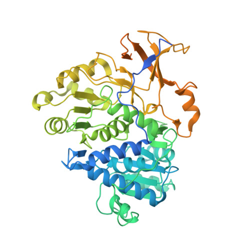
Crystal structure of native and a mutant of Lampyris turkestanicus luciferase implicate in bioluminescence color shift.
Kheirabadi, M., Sharafian, Z., Naderi-Manesh, H., Heineman, U., Gohlke, U., Hosseinkhani, S.(2013) Biochim Biophys Acta 1834: 2729-2735
- PubMed: 24103420 Search on PubMed
- DOI: https://doi.org/10.1016/j.bbapap.2013.09.022
- Primary Citation Related Structures:
3QYA, 4M46 - PubMed Abstract:
Firefly bioluminescence reaction in the presence of Mg(2+), ATP and molecular oxygen is carried out by luciferase. The luciferase structure alterations or modifications of assay conditions determine the bioluminescence color of firefly luciferase. Among different beetle luciferases, Phrixothrix hirtus railroad worm emits either yellow or red bioluminescence color. Sequence alignment analysis shows that the red-emitter luciferase from Phrixothrix hirtus has an additional arginine residue at 353 that is absent in other firefly luciferases. It was reported that insertion of Arg in an important flexible loop350-359 showed changes in bioluminescence color from green to red and the optimum temperature activity was also increased. To explain the color tuning mechanism of firefly luciferase, the structure of native and a mutant (E354R/356R/H431Y) of Lampyris turkestanicus luciferase is determined at 2.7Å and 2.2Å resolutions, respectively. The comparison of structure of both types of Lampyris turkestanicus luciferases reveals that the conformation of this flexible loop is significantly changed by addition of two Arg in this region. Moreover, its surface accessibility is affected considerably and some ionic bonds are made by addition of two positive charge residues. Furthermore, we noticed that the hydrogen bonding pattern of His431 with the flexible loop is changed by replacing this residue with Tyr at this position. Juxtaposition of a flexible loop (residues 351-359) in firefly luciferase and corresponding ionic and hydrogen bonds are essential for color emission.
- Department of Biophysics, Faculty of Biological Sciences, Tarbiat Modares University, Tehran 14115-175, Iran; Department of Biology, Faculty of Science, Hakim Sabzevari University, Khorasan razavi, Sabzevar 17976-487, Iran.
Organizational Affiliation: