


Enhanced and tunable fluorescent quantum dots within a single crystal of protein
Wei, H., House, S., Wu, J., Zhang, J., Wang, Z., He, Y., Gao, Y.-G., Robinson, H., Li, W., Zuo, J.-M., Robertson, I.M., Lu, Y.To be published.
Experimental Data Snapshot
wwPDB Validation 3D Report Full Report

Entity ID: 1 | |||||
|---|---|---|---|---|---|
| Molecule | Chains | Sequence Length | Organism | Details | Image |
| Lysozyme C | 129 | Gallus gallus | Mutation(s): 0 Gene Names: LYZ EC: 3.2.1.17 | 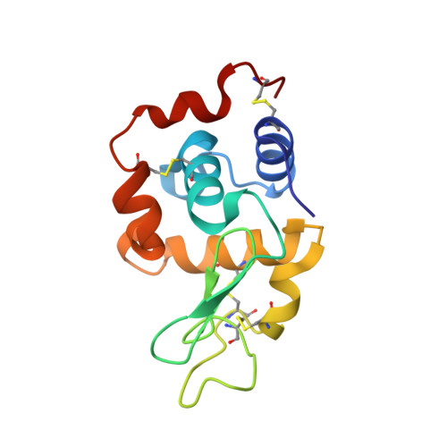 | |
UniProt | |||||
Entity Groups | |||||
| Sequence Clusters | 30% Identity50% Identity70% Identity90% Identity95% Identity100% Identity | ||||
| UniProt Group | P00698 | ||||
Sequence AnnotationsExpand | |||||
Reference Sequence | |||||
| Ligands 1 Unique | |||||
|---|---|---|---|---|---|
| ID | Chains | Name / Formula / InChI Key | 2D Diagram | 3D Interactions | |
| CD Download:Ideal Coordinates CCD File | B [auth A], C [auth A], D [auth A], E [auth A], F [auth A] | CADMIUM ION Cd WLZRMCYVCSSEQC-UHFFFAOYSA-N |  | ||
| Length ( Å ) | Angle ( ˚ ) |
|---|---|
| a = 78.677 | α = 90 |
| b = 78.677 | β = 90 |
| c = 37.251 | γ = 90 |
| Software Name | Purpose |
|---|---|
| SHELX | model building |
| SHELXL-97 | refinement |
| SHELX | phasing |


