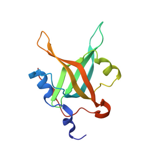
4LUV

Fragment-Based Discovery of a Potent Inhibitor of Replication Protein A Protein-Protein Interactions


Experimental Data Snapshot

  • Method: X-RAY DIFFRACTION
  • Resolution: 1.40 Å
  • R-Value Free: 0.170 
  • R-Value Work: 0.139 
  • R-Value Observed: 0.140 

Starting Model: experimental
View more details

wwPDB Validation   3D Report Full Report


Ligand Structure Quality Assessment 


This is version 1.2 of the entry. See complete history


Literature

Discovery of a potent inhibitor of replication protein a protein-protein interactions using a fragment-linking approach.

Frank, A.O.Feldkamp, M.D.Kennedy, J.P.Waterson, A.G.Pelz, N.F.Patrone, J.D.Vangamudi, B.Camper, D.V.Rossanese, O.W.Chazin, W.J.Fesik, S.W.

(2013) J Med Chem 56: 9242-9250

  • DOI: https://doi.org/10.1021/jm401333u
  • Primary Citation of Related Structures:  
    4LUO, 4LUV, 4LUZ, 4LW1, 4LWC, 4O0A

  • PubMed Abstract: 

    Replication protein A (RPA), the major eukaryotic single-stranded DNA (ssDNA)-binding protein, is involved in nearly all cellular DNA transactions. The RPA N-terminal domain (RPA70N) is a recruitment site for proteins involved in DNA-damage response and repair. Selective inhibition of these protein-protein interactions has the potential to inhibit the DNA-damage response and to sensitize cancer cells to DNA-damaging agents without affecting other functions of RPA. To discover a potent, selective inhibitor of the RPA70N protein-protein interactions to test this hypothesis, we used NMR spectroscopy to identify fragment hits that bind to two adjacent sites in the basic cleft of RPA70N. High-resolution X-ray crystal structures of RPA70N-ligand complexes revealed how these fragments bind to RPA and guided the design of linked compounds that simultaneously occupy both sites. We have synthesized linked molecules that bind to RPA70N with submicromolar affinity and minimal disruption of RPA's interaction with ssDNA.


  • Organizational Affiliation

    Department of Biochemistry, Vanderbilt University School of Medicine , Nashville, Tennessee 37232-0146, United States.


Macromolecules
Find similar proteins by:  (by identity cutoff)  |  3D Structure
Entity ID: 1
MoleculeChains Sequence LengthOrganismDetailsImage
Replication protein A 70 kDa DNA-binding subunit123Homo sapiensMutation(s): 1 
Gene Names: RPA1REPA1RPA70
UniProt & NIH Common Fund Data Resources
Find proteins for P27694 (Homo sapiens)
Explore P27694 
Go to UniProtKB:  P27694
PHAROS:  P27694
GTEx:  ENSG00000132383 
Entity Groups  
Sequence Clusters30% Identity50% Identity70% Identity90% Identity95% Identity100% Identity
UniProt GroupP27694
Sequence Annotations
Expand
  • Reference Sequence
Small Molecules
Ligands 2 Unique
IDChains Name / Formula / InChI Key2D Diagram3D Interactions
1DZ
Query on 1DZ

Download Ideal Coordinates CCD File 
C [auth A]1-(3-methylphenyl)-5-phenyl-1H-pyrazole-3-carboxylic acid
C17 H14 N2 O2
XDKDJRWOHMIRGN-UHFFFAOYSA-N
1XS
Query on 1XS

Download Ideal Coordinates CCD File 
B [auth A]5-(3-chloro-4-fluorophenyl)furan-2-carboxylic acid
C11 H6 Cl F O3
MGUVQRGVUWXOGD-UHFFFAOYSA-N
Binding Affinity Annotations 
IDSourceBinding Affinity
1XS PDBBind:  4LUV Kd: 1.40e+6 (nM) from 1 assay(s)
Experimental Data & Validation

Experimental Data

  • Method: X-RAY DIFFRACTION
  • Resolution: 1.40 Å
  • R-Value Free: 0.170 
  • R-Value Work: 0.139 
  • R-Value Observed: 0.140 
  • Space Group: P 21 21 21
Unit Cell:
Length ( Å )Angle ( ˚ )
a = 37.971α = 90
b = 53.528β = 90
c = 54.046γ = 90
Software Package:
Software NamePurpose
HKL-2000data collection
PHENIXmodel building
PHENIXrefinement
HKL-2000data reduction
HKL-2000data scaling
PHENIXphasing

Structure Validation

View Full Validation Report


Ligand Structure Quality Assessment 


Entry History 

Revision History  (Full details and data files)

  • Version 1.0: 2013-12-11
    Type: Initial release
  • Version 1.1: 2014-04-09
    Changes: Database references
  • Version 1.2: 2023-09-20
    Changes: Data collection, Database references, Derived calculations, Refinement description