4LRR | pdb_00004lrr

Ternary complex between E. coli thymidylate synthase, dUMP, and F9


Experimental Data Snapshot

  • Method: X-RAY DIFFRACTION
  • Resolution: 2.41 Å
  • R-Value Free: 
    0.230 (Depositor), 0.228 (DCC) 
  • R-Value Work: 
    0.170 (Depositor), 0.170 (DCC) 
  • R-Value Observed: 
    0.173 (Depositor) 

wwPDB Validation 3D Report Full Report

Validation slider image for 4LRR

Ligand Structure Quality Assessment 


This is version 1.2 of the entry. See complete history

Literature

2'-Deoxyuridine 5'-Monophosphate Substrate Displacement in Thymidylate Synthase through 6-Hydroxy-2H-naphtho[1,8-bc]furan-2-one Derivatives.

Ferrari, S.Calo, S.Leone, R.Luciani, R.Costantino, L.Sammak, S.Di Pisa, F.Pozzi, C.Mangani, S.Costi, M.P.

(2013) J Med Chem 56: 9356-9360

  • DOI: https://doi.org/10.1021/jm4014086
  • Primary Citation Related Structures: 
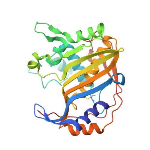
    4LRR

  • PubMed Abstract: 

    Thymidylate synthase (TS) is a target for antifolate-based chemotherapies of microbial and human diseases. Here, ligand-based, synthetic, and X-ray crystallography studies led to the discovery of 6-(3-cyanobenzoyloxy)-2-oxo-2H-naphto[1,8-bc]furan, a novel inhibitor with a Ki of 310 nM against Pneumocystis carinii TS. The X-ray ternary complex with Escherichia coli TS revealed, for the first time, displacement of the substrate toward the dimeric protein interface, thus providing new opportunities for further design of specific inhibitors of microbial pathogens.


  • Organizational Affiliation
    • Department of Life Sciences, University of Modena and Reggio Emilia , Via Campi 183, 41125 Modena, Italy.

Macromolecule Content 

  • Total Structure Weight: 32 kDa 
  • Atom Count: 2,395 
  • Modeled Residue Count: 265 
  • Deposited Residue Count: 265 
  • Unique protein chains: 1

Macromolecules

Find similar proteins by:|  3D Structure
Entity ID: 1
MoleculeChains  Sequence LengthOrganismDetailsImage
Thymidylate synthase265Escherichia coli O157:H7Mutation(s): 1 
Gene Names: thyA
EC: 2.1.1.45
UniProt
Find proteins for P0A886 (Escherichia coli O157:H7)
Explore P0A886 
Go to UniProtKB:  P0A886
Entity Groups
Sequence Clusters30% Identity50% Identity70% Identity90% Identity95% Identity100% Identity
UniProt GroupP0A886
Sequence Annotations
Expand
Reference Sequence

Small Molecules

Ligands 3 Unique
IDChains Name / Formula / InChI Key2D Diagram3D Interactions
CF9

Query on CF9


Download:Ideal Coordinates CCD File
J [auth A]2-oxo-2H-naphtho[1,8-bc]furan-6-yl 4-nitrobenzoate
C18 H9 N O6
JZRRRFGHBXIXDI-UHFFFAOYSA-N
UMP

Query on UMP


Download:Ideal Coordinates CCD File
K [auth A]2'-DEOXYURIDINE 5'-MONOPHOSPHATE
C9 H13 N2 O8 P
JSRLJPSBLDHEIO-SHYZEUOFSA-N
SO4

Query on SO4


Download:Ideal Coordinates CCD File
B [auth A]
C [auth A]
D [auth A]
E [auth A]
F [auth A]
B [auth A],
C [auth A],
D [auth A],
E [auth A],
F [auth A],
G [auth A],
H [auth A],
I [auth A]
SULFATE ION
O4 S
QAOWNCQODCNURD-UHFFFAOYSA-L
Modified Residues  1 Unique
IDChains TypeFormula2D DiagramParent
SCH
Query on SCH
A
L-PEPTIDE LINKINGC4 H9 N O2 S2CYS

Experimental Data & Validation

Experimental Data

  • Method: X-RAY DIFFRACTION
  • Resolution: 2.41 Å
  • R-Value Free:  0.230 (Depositor), 0.228 (DCC) 
  • R-Value Work:  0.170 (Depositor), 0.170 (DCC) 
  • R-Value Observed: 0.173 (Depositor) 
Space Group: I 21 3
Unit Cell:
Length ( Å )Angle ( ˚ )
a = 131.184α = 90
b = 131.184β = 90
c = 131.184γ = 90
Software Package:
Software NamePurpose
MOLREPphasing
REFMACrefinement
MOSFLMdata reduction
SCALAdata scaling

Structure Validation

View Full Validation Report


Ligand Structure Quality Assessment 


Entry History 

Deposition Data

Revision History  (Full details and data files)

  • Version 1.0: 2013-11-06
    Type: Initial release
  • Version 1.1: 2013-12-11
    Changes: Database references
  • Version 1.2: 2025-03-26
    Changes: Data collection, Database references, Derived calculations, Structure summary